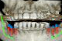
Cone-beam CT (CBCT) images can help assess the loss of periodontal tissue and classify the degree of furcation involvement in maxillary molars, according to researchers from the University of Basel (Journal of Clinical Periodontology, May 2010, Vol. 37:5, pp. 436-441).
For their study, 14 patients with generalized advanced chronic periodontitis were consecutively recruited and treated nonsurgically. CBCT was performed on maxillary molars considered for furcation surgery due to increased furcation involvement (FI) and/or increased probing pocket depths during re-evaluation, and the degree of involvement was evaluated from the images.
Furcation surgery was performed in 25 maxillary molars. Intrasurgical FI assessments were compared with data derived from CBCT images.
Overall, 84% of the CBCT data were confirmed by the intrasurgical findings. While 14.7% (11 sites) were underestimated (cone-beam CT less than intrasurgical value), in only one site did the CBCT data lead to an overestimation compared with the intrasurgical analysis.
"Cone-beam CT images demonstrate a high accuracy in assessing the loss of periodontal tissue and classifying the degree of furcation involvement in maxillary molars," the authors concluded.
Copyright © 2010 DrBicuspid.com