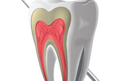
Researchers have shown that two regenerative endodontic procedures (REPs) -- revitalization and a platelet-rich, fibrin (PRF)-based technique -- could be a viable treatment for mature permanent teeth with necrotic pulps. Their findings were published recently in the International Endodontic Journal.
Traditionally, necrotic pulps have been treated with root canal therapy. But the results of the randomized, prospective, controlled trial in Egypt demonstrated the success of both regenerative endodontic procedures.
"Our findings indicated that REPs using the [blood clot] technique or PRF technique can be a treatment option for mature teeth with a necrotic pulp," the authors wrote, led by Ahmed Youssef of the endodontic department at Minia University in Egypt (Int Endod J, January 14, 2022).
The potential of regenerative treatment
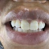
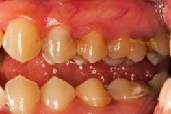
To assess the potential of regenerative endodontic procedures, the researchers randomly selected 20 patients with mature necrotic anterior teeth with large periapical lesions from among those seeking treatment at the endodontic outpatient clinic associated with Minia University.
The patients, who ranged in age from 18 to 40, were randomly assigned to two groups of 10 for two different REP treatments. The first group was treated with revitalization using the blood clot technique, and the second group was treated using the PRF-based technique.
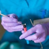
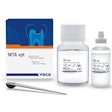
In the blood clot group, bleeding was evoked in the canal and controlled at a level just below the cementoenamel junction. The canals were left for seven minutes to allow a blood clot to form. White mineral trioxide aggregate (MTA) was then placed approximately 3 mm below the junction, followed by a moist cotton pellet and a temporary filling to allow for complete setting of the MTA. After two days, the temporary filling was replaced by a glass-ionomer cement base and resin composite restoration to seal the access cavity.
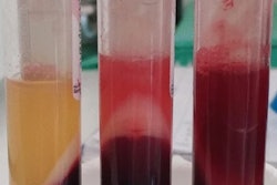
In the PRF group, PRF was prepared by drawing blood from the patient's forearm into a test tube without an anticoagulant. The blood was centrifuged, and the PRF was segregated and squeezed to form a membrane. Similar to the blood clot group, bleeding was evoked in the canal, and the PRF membrane was fragmented and placed incrementally in the canal up to the level of the cementoenamel junction. Then a 3-mm-thick layer of white MTA was placed directly over the PRF matrix.
After treatment, the patients' periradicular healing was assessed using standardized radiographs taken at baseline with a flat-bed scanner at six and 12 months after treatment. Each patient's periapical status was assessed by multiple observers using the periapical index score. An electric pulp tester was also used to assess whether pulp sensibility had been regained.
The results showed a significant increase in periradicular healing in both groups at six and 12 months compared to the patients' status at baseline. Neither group outperformed the other, and there was no significant difference in the periapical index scores between the two groups after 12 months.
When pulp sensibility was assessed after one year of follow-up, two patients in the blood clot group and five patients in the PRF group were found to have regained sensibility. The researchers speculated that the relative success of the PRF group may have been due to the different growth factors available in PRF, including those important for neurogenesis.
"The findings of this preliminary trial indicate the potential for using REPs, such as revitalization or PRF-based techniques, as treatment options for mature teeth with necrotic pulp," the authors concluded. "A higher level of evidence is required however, with appropriately sized prospective clinical trials and longer follow ups to conclusively validate the different outcomes of REPs."
