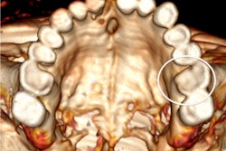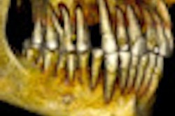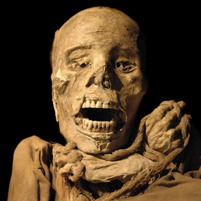
Analysis of pre-Hispanic mummies found in the Canary Islands has shown that their dental pathologies did not differ significantly from skeletonized remains found on the same island, according to research published online July 13 in the International Journal of Paleopathology.
A research group performed macroscopic examination under fluorescent lights of pathological and nonpathological features of the oral cavity in 400 teeth and 764 alveoli of 30 adult pre-Hispanic mummies from the fifth to 11th centuries A.D. on Gran Canaria, the third largest of the Canary Islands. The team, led by Teresa Delgado-Darias of the archaeological museum El Museo Canario in Gran Canaria, concluded that the dental pathology profile of the mummies indicated a carbohydrate-rich diet that contained abrasive grit from the stone querns used to grind cereals.
Nearly 12% of the mummies had dental caries, of which 28% affected the molars. About two-thirds of the mummies had a high prevalence of calculus, and roughly another one-third exhibited signs of periodontal disease. Other findings included periapical lesions in 11% of the mummies and antemortem tooth loss in 16%.
The authors described the wear as "extensive dentine exposure." In total, 84% of the mummies showed signs of linear enamel hypoplasia.
The researchers found no statistically significant differences, however, when they compared their findings with previously published data from skeletonized remains found on the same island. The results reinforce the recent rejection of the traditional interpretation of the mummies as the preeminent status group of Canarian society, according to the researchers.
"This is the first study to delve into the oral conditions of pre-Hispanic mummified remains from Gran Canaria," the authors wrote. "The results have implications for the framing of research questions based on the social status of these mummies."


