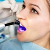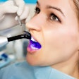
Hard-tissue lasers have been praised as a replacement to drills for basic restorative procedures, which is why researchers decided to take a closer look at how one type, the erbium-doped yttrium aluminium garnet (Er:YAG) laser, affected dentin's physical and chemical properties.
The Er:YAG laser increased dentin surface hardness but decreased the calcium-phosphorous ratio compared with conventional drills, the researchers found. Their findings were published in Lasers in Medical Science (August 1, 2017).
"[The] Er:YAG laser has been used for caries removal in permanent and primary teeth, eliminating the vibration, pressure, and noise during tooth preparation [and] being preferable and well accepted by patients because it eliminates the need for local anesthesia in most cases," wrote the authors, led by Fabiana Curylofo-Zotti, BDS, from the University of São Paulo School of Dentistry in Brazil. "Despite the ablation effectiveness, the structural and chemical aspects of the demineralized dentin irradiated with Er:YAG laser were not clarified."
Er:YAG laser vs. conventional drill
Hard-tissue lasers, including Er:YAG lasers, have been touted as a comfortable and quiet way to remove caries in primary and permanent teeth, as well as to address hypersensitivity. However, some studies have suggested that the lasers can also demineralize dentin and kill pulp. The researchers, therefore, wanted to see how Er:YAG lasers affected the hardness and chemical composition of dentin.
To find out, they crafted 104 bovine incisors into 5 x 5-mm dentin slabs. The researchers specifically chose cattle teeth because of their similarity to human teeth in hardness and chemical composition.
They began their experiment by cleaning and polishing the dentin slabs before taking initial microhardness measurements using a microhardness tester, the HMV-2000 (Shimadzu), using Knoop hardness values. They took the measurements 30 µm from the surface with 100 µm of space between them.
Once the initial measurements were taken, the researchers ran the slabs through pH cycling to create caries. They then applied two layers of acid-resistant varnish to all but one of the dentin slab sides.
The slabs were then left in 10 mL of demineralizing solution for eight hours, followed 10 mL of remineralizing solution for 16 hours. The researchers repeated the demineralization and remineralization cycles every day for 14 days and took microhardness measurements for a second time at the end of the two-week cycle.
The slabs were then divided into two groups. For the first group, the researchers removed caries with a low-speed, spherical carbide drill as a control. For the second, they removed caries using an Er:YAG laser (Fidelis Plus III, Fotona) with the following settings:
- Focal distance of 7 mm
- Pulse energy output of 250 millijoule (mJ)
- Pulse rate of 4 hertz
- Beam diameter of 0.99 mm
- Energy density of 39 J/cm2
With the carious lesions removed, the researchers once again performed the microhardness tests before filling in the restoration with an adhesive and composite resin (Filtek Z350, 3M) and taking final microhardness measurements.
| Dentin microhardness (Knoop hardness values) |
||
| Bur | Er:YAG | |
| Before caries induction | 52.8 | 55.3 |
| After caries induction | 15.7 | 14.4 |
| After treatment and restoration | 22.1 | 33.4 |
Dentin microhardness was significantly higher for surfaces treated with the Er:YAG laser compared with the drill, the researchers found.
“The removal of caries with Er:YAG laser increased the microhardness of the residual caries-affected dentin.”
After further studying the dentin slabs with scanning electron microscopy, the researchers noted the tooth surfaces treated with Er:YAG were scaly with visible tubule orifices, while a smear layer was present on those treated with the bur. These surface changes may be responsible for the difference in hardness between the two groups, they noted.
"In the present study, the removal of caries with Er:YAG laser increased the microhardness of the residual caries-affected dentin compared to bur at low-speed handpiece," the authors wrote. "An increase in microhardness values of dentin is related to an increase of the hardness of intertubular dentin."
However, surfaces treated with the Er:YAG laser also showed a significant decrease in the amount of calcium, phosphorous, and the calcium-phosphorous ratio, which may negatively affect the ability of restorative material to adhere to the treated tooth surfaces. The researchers attributed this to chemical changes prompted by the laser.
"Changes in calcium/phosphorous ratio indicate that the original relationship between the organic and inorganic components was changed," they wrote. "This may affect the permeability, solubility, or adhesive pattern of the dental hard tissues."
Innovations needed
The researchers noted that their findings differed from previous studies that were similar. They chalked up the differing results to variances in how they prepared the cavities, when hardness measurements were taken, and the settings used for both the drills and the lasers.
The researchers also suggested a need for an agent that can repair the quantities of calcium ions and the calcium/phosphorous ratio. In the study, they tried chitosan as a modification, but it did not have a significant difference on the hardness or calcium-phosphate levels.
"Thus, it can be concluded that the selective removal of carious dentin with Er:YAG laser increased microhardness of residual caries-affected dentin changing its surface morphology and chemical composition," the authors wrote. "The biomodification with chitosan did not influence the structural and chemical composition of residual caries-affected dentin."
