Can the addition of a calcium phosphate to a glass ionomer cement (GIC) decrease the amount of plaque-causing biofilms and reduce caries?
In the first study in which glass ionomer cement disks have been imaged undisturbed and in real-time, researchers from Australia examined what would happen to biofilms when 3% casein phosphopeptide-amorphous calcium phosphate (CPP-ACP) was added to these disks.
"It was proposed that by adding CPP-ACP to this GIC there would be a greater inhibition of bacterial binding and proliferation conferring greater protection of the tooth surface at risk of developing caries," wrote study author Stuart Dashper, PhD, and colleagues from the Oral Health Cooperative Research Centre at the Melbourne Dental School (PLOS One, September 2, 2016).
Reducing damage
The researchers sought to see if there was a way to reduce bacteria-associated damage to the surface of glass ionomer cement and also reduce the risk of caries development. Streptoccus mutans is considered a major agent of caries due to its "aciduric and highly acidogenic nature," the authors wrote.
“In the clinical setting, the use of CPP-ACP containing GIC coupled with regular aqueous CPP-ACP treatment may reduce bacterial biofilm establishment.”
Glass ionomer cements have been referred to as "smart materials," because some of their properties can be changed in response to conditions. One such change is using 3% CPP-ACP to inhibit enamel demineralization and allow calcium and phosphate ions to inhibit S. mutans' effects.
CPP-ACP has been shown in previous studies to both slow caries progression of caries and allow enamel subsurface lesions to remineralize. The researchers wanted to see how adding CPP-ACP to a GIC mixture would affect the development of S. mutans biofilms.
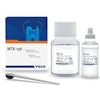
They used Fuji VII and Fuji VII EP glass ionomer cement (GC) containing 3% weight/weight (w/w) CPP-ACP. The S. mutans biofilms were either established in flow cells before a 10-minute exposure to 1% weight/volume (w/v) CPP-ACP or cultured in static wells or flow cells with either GIC or GIC containing 3% w/w CPP-ACP. The researchers used laser scanning microscopy after staining to visualize the biofilms.
The incorporation of 3% CPP-ACP into the GIC significantly inhibited S. mutans biofilm development by more than 50% (see table below), they found.
| Biometric parameters of 16-hour static S. mutans biofilms | |||
| Fuji VII GIC (no CPP-ACP) |
Fuji VII EP (3% CPP-ACP) |
% change | |
| Biovolume (µm3/µm2) | 1.53 ± 0.33 | 0.76 ± 0.23* | -50% |
| Average thickness (µm) | 1.88 ± 0.61 | 0.65 ± 0.11* | -66% |
| Maximum thickness (µm) | 21.56 ± 8.94 | 17.16 ± 3.78 | -19% |
| Surface area: Volume ratio | 2.27 ± 0.45 | 3.24 ± 0.63* | +70% |
| Proportion of the substratum colonized (%) | 14.92 ± 5.07 | 12.85 ± 4.50 | -14% |
This treatment also significantly altered the structure of the biofilms compared with controls, the researchers found. This suggests that it was biofilm development that was inhibited by CPP-ACP that had diffused from the GIC.
Biofilm development
The authors noted that their results were consistent with other studies on GICs’ inherent inhibitory effect on bacterial growth. They speculated that this inhibitory effect may be caused by the release of fluoride or other ions.
"In the clinical setting, the use of CPP-ACP containing GIC coupled with regular aqueous CPP-ACP treatment may reduce bacterial biofilm establishment and development and, therefore, may reduce bacteria-associated damage to the GIC surface and the development of caries," the authors concluded.
