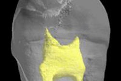
Stem cells from the dental pulp of third molars can be transformed into cells of the eye's cornea and could one day be used to repair corneal scarring due to infection or injury, according to a new study in Stem Cells Translational Medicine (February 23, 2015).
The findings indicate these stem cells also could become a new source of corneal transplant tissue made from the patient's own cells.
Corneal blindness, which affects millions of people worldwide, is typically treated with transplants of donor corneas, according to senior investigator James Funderburgh, PhD, professor of ophthalmology at the University of Pittsburgh (Pitt) and associate director of the Louis J. Fox Center for Vision Restoration of the University of Pittsburgh Medical Center.
"Shortages of donor corneas and rejection of donor tissue do occur, which can result in permanent vision loss," Funderburgh said in a statement. "Our work is promising because using the patient's own cells for treatment could help us avoid these problems."
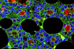
Experiments conducted by lead author Fatima Syed-Picard, PhD, also of Pitt's department of ophthalmology, showed that stem cells of dental pulp, obtained from routine human third molar extractions performed at Pitt's School of Dental Medicine, could be turned into corneal stromal cells called keratocytes, which have the same embryonic origin.
The team injected the engineered keratocytes into the corneas of healthy mice, where they integrated without signs of rejection. They also used the cells to develop constructs of corneal stroma akin to natural tissue.
"Other research has shown that dental pulp stem cells can be used to make neural, bone, and other cells," Syed-Picard noted. "They have great potential for use in regenerative therapies."
In future work, the researchers will assess whether the technique can correct corneal scarring in an animal model.
The project was funded National Institutes of Health grants and the Eye and Ear Foundation of Pittsburgh.
