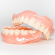
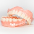
A man unknowingly swallowed his partial denture, causing an abnormal connection between his esophagus and trachea that required him to undergo two surgeries and eight months of recovery. The case report was recently published in the Journal of Cardiothoracic Surgery.
Surgical management of an acquired, nonmalignant tracheoesophageal fistula (TOF) secondary to denture impaction is not only very rare, but it creates a clinical challenge. After the surgeries, the man dealt with repeated physiological problems, permanent loss of phonation, and negative psychological effects. He had to build his stamina to eat and drink a variety of foods and liquids and he had to use an electrolarynx device to speak, the authors wrote.
"In summary, we have reported on a complex case of an acquired, non-malignant TOF secondary to chronically impacted denture which is rarely described in the literature,” wrote the authors, led by Dr. Hannah Jesani of the University Hospitals Birmingham in the United Kingdom (J Cardiothorac Surg, November 4, 2024, Vol. 19, 621).
A man in his 60s with dysphagia, chest infections
A man visited a hospital emergency department for complaints of coughing and regurgitation after eating. He had a history of trouble swallowing solids and liquids, weight loss, and recurring chest infections that were ongoing for more than a year. His symptoms worsened two weeks before he went to the hospital, according to the case report.
The patient reported wearing a dental prosthesis but did not recall swallowing it. His only medical history was being diagnosed with epilepsy, and he did not smoke, drank alcohol occasionally, and was otherwise independent with daily activities, the authors wrote.
He underwent a barium swallow fluoroscopy that revealed an irregularity of the esophageal tract, aspiration of contrast on swallowing, and findings of an esophageal mass. A computed tomography (CT) scan of the man's neck, thorax, abdomen, and pelvis with contrast reported esophageal wall thickening, suggesting a fistula between the esophagus and proximal trachea. Also, there was shadowing indicative of an infection, they wrote.
 Preoperative barium swallow fluoroscopy study. Shows contrast in the trachea and esophagus (red arrows), suggesting a fistulous communication. Images and captions courtesy of Dr. Hannah Jesani, et al. Licensed under CC BY 4.0.
An endoscopic procedure revealed difficult visualization, which showed a "large, stalked polyplike" lesion, which made it difficult to biopsy. After further inspection of the imaging, he was diagnosed with a foreign object located in the fistulous tract.
 Axial slices of the computed tomography scan with contrast. Images show a linear hyperdense shadow extending from the esophagus to the proximal trachea within a tracheo-esophageal fistulous tract at the level of T2 vertebrae.
Doctors attempted to remove the object endoscopically, but the procedure was abandoned once it was determined that it was a denture with sharp edges. Doctors feared that trying to remove the deeply impacted denture could result in bleeding or further injuries to the man's trachea or esophagus. Therefore, an open surgical procedure was performed to remove the denture, and he was given a feeding tube, the authors wrote.
Following the procedure, he experienced many complications, including vocal cord palsy and trouble weaning from ventilation. He spent 62 days in the intensive care unit, they wrote.
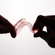
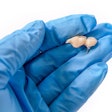
 The extracted foreign body: a removable, partial denture.
The man underwent a pharyngolaryngectomy, permanent tracheostomy, and restoration of the digestive tract. His recovery and rehabilitation, which included speech and language therapy, nutritional counseling, and more occurred over eight months.
What to remember
This case highlights the importance of a collaborative medical strategy when denture ingestion or impaction occurs. In this instance, extensive preparation, postoperative rehabilitation, and surgical expertise were necessary to repair the fistula and restore the patient's digestive tract, the authors wrote.
Recognizing an impacted foreign body is crucial to successful treatment. Prolonged impaction in the esophagus can lead to a pathological process of edema, infection, and necrosis, they wrote.
"Early recognition and removal of impacted dental prosthesis is essential to prevent morbidity and mortality," Jesani and colleagues wrote.


