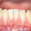
By using fluorodeoxyglucose positron emission tomography (FDG-PET) to measure metabolic activity, researchers at Massachusetts General Hospital and Harvard Medical School have found a link between periodontal disease and atherosclerotic inflammation, according to a study in the Journal of the American College of Cardiology (February 22, 2011, Vol. 57:8, pp. 971-976).
In their 112-patient study, the researchers measured periodontal FDG uptake by obtaining standardized uptake values (SUVs) from the periodontal tissue of each patient. They also calculated the ratio of periodontal activity to background (blood) activity (TBR).
SUV measurements were also obtained in the carotid and aorta, as well as in a venous structure. Localization of periodontal, carotid, and aortic activity was facilitated by PET coregistration with CT or MRI.
A subset of 16 patients underwent carotid endarterectomywithin one month of PET imaging, during which atherosclerotic plaques were removed and subsequently stained with antibodies to quantify macrophage infiltration. Periodontal FDG uptake was compared with carotid plaque macrophage infiltration.
The analysis revealed an association between periodontal FDG uptake (TBR) and carotid TBR and aortic TBR. In addition, the researchers found a "strong relationship" between periodontal TBR and histologically assessed inflammation within carotid artery plaques.
"These findings provide direct evidence for an association between periodontal disease and atherosclerotic inflammation," wrote lead study author Kenneth Fifer, from the university's division of cardiology and cardiac MR-PET-CT program, and colleagues.


