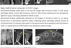
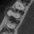
Cone-beam CT (CBCT) can be used to better determine root canal length and width, improving the prediction of root canal treatment outcomes, according to a study in Oral Medicine, Oral Pathology, and Oral Surgery (August 15, 2010).
While many studies have investigated the histological changes in pulp, few have focused on the changes in the shape of the root canals, according to researchers from Dicle University in Turkey.
They used cone-beam CT to image 100 noncarious maxillary central teeth. These teeth were divided into five groups according to the age of the patients: group A:15-24, group B: 25-34, group C: 35-44, group D: 45-54, and group E: 55 years and older. The cone-beam CT scans were used to determine root length and pulp width at the cervical, apical 1/2, and apical 1/3.
On comparing the groups using one-way analysis of variance (ANOVA), the researchers found that the root length did not change with aging (p > 0.05), while the pulp width at the cervical, apical 1/2, and apical 1/3 differed among the groups (p < 0.001).
"Cone-beam CT can be used to determine the precise root canal length and width ... and prevent iatrogenic exposure of the apex," the researchers wrote. "This will improve the prediction of the prognosis of root canal treatment."
Copyright © 2010 DrBicuspid.com

