Cone-beam CT (CBCT) scans are significantly more efficient than periapical radiographs in determining the mesiobuccal root canal anatomy of maxillary molars, according to a study to be presented at the International Association for Dental Research meeting in July in Barcelona, Spain.
Using a new in vitro model of dentoalveolar structure suitable for radiographic imaging, researchers from the University of California, Los Angeles coated 50 extracted maxillary first molars with a thin layer of dental wax to simulate the periodontal ligament. They also placed a mixture of calcium phosphate and petroleum jelly, which mimics normal bone density on radiographs, on the teeth models.
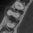
Three periapical views (straight-on, mesial, and distal) of the models were taken, along with CBCT scans. Some were also imaged using micro-CT as a positive control. The CBCT images were further studied to determine the microanatomical features, including the location of the second mesiobuccual canal in relation to other structures, classification of the mesiobuccal root canal system, presence of the isthmus, and location of portals of exit.
The periapical images identified two teeth with second mesiobuccual canals but were unable to determine their distance from the mesiobuccal canal. In contrast, CBCT images were able to locate second mesiobuccual canals in all teeth evaluated, with an average distance of 2.056 mm at the orifice level, 2.792 mm at the furcation level, and 2.231 mm at the midroot level to mesomuccal canal.
From these results, the researchers concluded that CBCT scans "will aid in enhancing the endodontic clinical outcome."
Copyright © 2010 DrBicuspid.com

