A team of Indian researchers led by Narayanan Subhash, PhD, has developed a noninvasive spectral imaging system that could provide rapid, inexpensive mass screening for oral cancers in dental and clinical settings, according to a press release by Andor Technology.
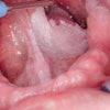
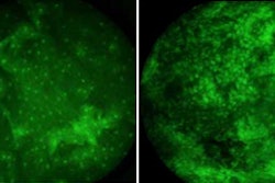
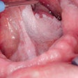
The core of their diffuse reflectance imaging system (DRIS) is the Andor Luca-R EMCCD camera, which captures monochrome images of the patient's mouth at 545 nm and 575 nm, according to the company.
Andor's Solis software computes a ratio image (R545/R575) of the area under investigation and generates a pseudo color map (PCM) where blue designates healthy tissue, red denotes dysplastic/premalignant tissue, and yellow identifies malignant tissue. This allows rapid visual differentiation of oral lesions and identification of regions with premalignant characteristics.
"[Our imaging method] delineates the boundaries of neoplastic changes and locates sites with the most malignant potential for biopsy, thereby avoiding unnecessary repeated biopsies and delay in diagnosis," s Subhash stated in the press release. "What's more, imaging the entire region may also help the surgeons to identify the margins of the lesion that cannot be easily visualized by the naked eye during surgical interventions."


