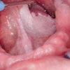
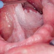
Researchers from Queen Mary, University of London have developed a new gene test that may detect precancerous cells in patients with oral lesions that appear to be benign.
A study in the International Journal of Cancer (October 4, 2012) showed that the quantitative Malignancy Index Diagnostic System (qMIDS) test had a cancer detection rate of 91% to 94% when used on more than 350 head and neck tissue specimens from 299 patients in the U.K. and Norway.
"A sensitive test capable of quantifying a patient's cancer risk is needed to avoid the adoption of a ‘wait-and-see' intervention," stated lead investigator and inventor of the test Muy-Teck Teh, PhD, in a university press release. "Detecting cancer early, coupled with appropriate treatment can significantly improve patient outcomes, reduce mortality, and alleviate long-term public healthcare costs."
The qMIDS test measures the levels of 16 genes that are converted, via a diagnostic algorithm, into a "malignancy index" which quantifies the risk of the lesion becoming cancerous. It is less invasive than the standard histopathology methods as it requires a 1- to 2-mm piece of tissue (less than half a grain of rice), and it takes less than three hours to get the results, compared with up to a week for standard histopathology.
While this proof-of-concept study validates qMIDS as a diagnostic test for early cancer detection, further clinical trials are required to evaluate the long-term clinical benefits of the test for oral cancers.
With further development, it could potentially be applied to other cancer types as the test is based on a cancer gene -- FOXM1 -- which is highly expressed in many cancer types. In this study, the researchers used the qMIDS test to detect early cancer cells in vulva and skin specimens with promising results.
This study was jointly funded by the Facial Surgery Research Foundation - Saving Faces (U.K.), Bergen Medical Research Foundation, Norwegian Cancer Research Association, British Skin Foundation, and Cancer Research UK.


