Optical coherence tomography (OCT) offers a noninvasive approach for localizing the most representative site to biopsy in a suspicious oral lesion, according to a study in Lasers in Medical Science (August 18, 2011).
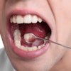
Selecting the most representative site for biopsy is crucial in establishing a definitive diagnosis of oral epithelial dysplasia, noted the study authors from Queens Mary University London.
Because current methods of clinical examination can result in sampling errors, the researchers tested OCT for differentiating normal and dysplastic oral epithelial samples and developing an objective and reproducible approach for biopsy site selection.
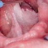
Biopsy samples from patients with fibroepithelial polyps (n = 13), mild dysplasia (n = 2), and moderate/severe dysplasia (n = 4) were scanned at 5-µm intervals using an OCT microscope and subsequently processed and stained with hematoxylin and eosin.
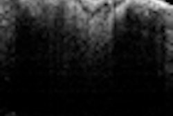
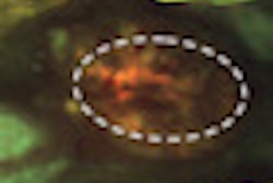
They found that the OCT images of normal oral epithelium showed a clear delineation of the mucosal layers observed in the matching histology. However, OCT images of oral dysplasia did not clearly identify the individual mucosal layers because of the increased density of abnormal cell nuclei, which impeded light penetration, the researchers noted.
"Quantitative differentiation of normal and dysplastic lesions using OCT offers a noninvasive objective approach for localizing the most representative site to biopsy, particularly in oral lesions with similar clinical features," the study authors concluded.