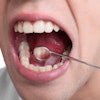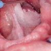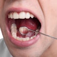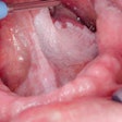
With the incidence of oral cancer on the rise worldwide -- nearly 130,000 deaths are attributed to this disease annually, according to the American Cancer Society -- there is a growing need to develop better methods for detecting it at an earlier stage.
The situation is even more critical in developing countries, according to researchers from Rice University and the M. D. Anderson Cancer Center who have been developing a new imaging device they believe will help address the problem. More than two-thirds of cases and three-quarters of deaths from oral cancer occur in developing countries, they noted in a study published in Head & Neck Oncology (April 22, 2010). In addition, while the overall five-year survival for patients with oral cancer in the U.S. is only 54%, five-year survival rates in developing countries are even worse: less than 30%.
“You have to have a battery-powered, very low-cost device — in the $100 to $200 range.”
— Rebecca Richards-Kortum, Ph.D.,
Rice University
"No satisfactory mechanism exists currently to screen and detect early neoplastic changes of the oral cavity in the general population," the researchers wrote. "The situation is even more challenging in developing countries and low-resource regions with high-risk populations, where a combination of lack of public awareness of the disease and inadequate resources and expertise for screening can result in even greater delays in diagnosis, leading to higher morbidity and mortality."
Oral cancer screening typically involves visual inspection of the entire tissue surface at risk under white-light illumination. But a number of products designed to improve diagnostic outcomes have been developed and commercialized, including the VELscope, Identafi, Vizilite Plus, Microlux, and OralCDx. And researchers continue to experiment with new ways to more accurately find and distinguish potentially malignant lesions using advanced optical technologies such as fluorescence spectroscopy, autofluorescence visualization, confocal microscopy, and optical coherence tomography.
Limited clinical resources
But developing countries pose unique challenges when it comes to creating a device that is affordable and can be used by individuals with limited clinical expertise.
"Our goal is to provide cost-effective opportunities that will work with the persons available in those health systems," said Rebecca Richards-Kortum, Ph.D., a professor of bioengineering at Rice University and lead author on the Head & Neck Oncology study. "What that means is that you have to have a battery-powered, very low-cost device -- in the $100 to $200 range. We are not there yet, but we are working on it. And it should have some type of objective image interpretation because there is a big shortage of skilled healthcare workers in these countries."
Richards-Kortum and her colleagues have spent that past decade developing optical imaging devices for cancer screening and diagnosis. For the Head & Neck Oncology study, which was conducted at Tata Memorial Hospital in Mumbai, India, they used a multimodal imaging system that incorporates blue and white LEDs to illuminate the oral mucosa. Images can be observed visually or captured digitally through a set of optical filters connected to a CCD camera. The system weighs only 3 lb and is powered by a lithium-ion battery.
In vivo images
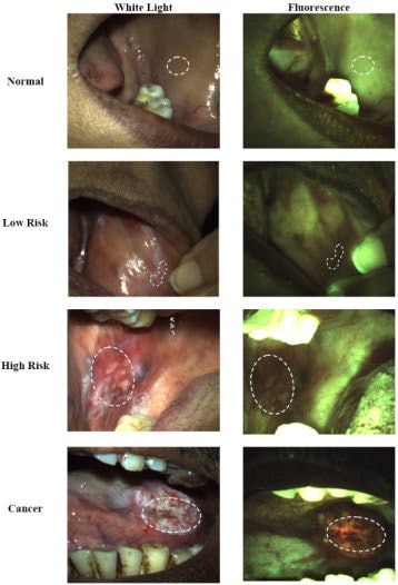 |
| Lesions typical of the four consensus clinical impression categories, with white light images on the left and fluorescence images on the right. Top row: Normal buccal mucosa, illustrating homogenous green fluorescence. Second row: Low-risk lesion of the buccal mucosa, with loss of fluorescence. Third row: High-risk lesion in the buccal mucosa, with loss of fluorescence. Bottom row: Rancer of the tongue, with loss of green fluorescence and presence of orange-red fluorescence. Rahman et al. Head & Neck Oncology 2010 2:10 doi:10.1186/1758-3284-2-10. |
The study included 76 patients who were referred to the Cancer Prevention Clinic at Tata Memorial Hospital because of suspicious oral lesions or who were waiting for head and neck surgery in the hospital, along with 33 healthy volunteers with and without a history of tobacco use.
Each patient was given a conventional exam of the oral cavity by a head and neck specialist. Initial clinical impression of each site as normal or abnormal was noted. In addition, the presence of either melanosis or oral submucous fibrosis was noted if visible. After clinical examination, digital reflectance and fluorescence images were obtained from clinically abnormal sites and contralateral clinically normal sites. Images were also obtained from the lateral border of the tongue, buccal mucosa, and lip of each subject whenever these sites were accessible.
Images from a total of 351 different sites were included in the analysis; 261 of these sites were imaged from 76 patients, and 90 sites were imaged from 33 normal volunteers. A total of 222 sites had a consensus clinical impression of normal, 30 sites had a consensus clinical impression of low risk for neoplasia, 22 sites had a consensus clinical impression of high risk for neoplasia, and 37 had a consensus clinical impression of cancer.
Good sensitivity and specificity
The system's image-analysis algorithm identified tissues judged clinically to be cancer or clinically suspicious for neoplasia with a sensitivity of 90% and a specificity of 87%, the researchers found.
"Although further work is needed to address the limitations in this study, results from this pilot study suggest that this simple imaging device can potentially improve oral screening efforts in low-resource settings where clinical expertise and resources are often limited," they concluded.
"This is a good development for oral cancer detection," said Mary Reid, Ph.D., a research scientist in the Division of Cancer Prevention and Population Sciences at Roswell Park Cancer Institute whose group has been working with autofluorescence-guided visualization to enhance oral cancer detection. "Our device is a little cumbersome. But theirs [Rice University's] looks a little easier to use. It is the kind of technology that is important, especially because this is a multifocal disease. Being able to identify high-risk groups is significant."
The Rice University results also compare favorably to those of other optical imaging systems. A review in General Dentistry (January 2009, Vol. 57:1, pp. 34-38) summarized several clinical studies of optical imaging with qualitative image analysis for detecting oral neoplasia and reported sensitivity of 63% to 100% and specificity of 79% to 96%. And a study in Cancer Prevention Research (May 2009, Vol. 2:5, pp. 423-431) reported sensitivity of 94% and specificity of 87% using quantitative analysis of fluorescence and reflectance images obtained with a multispectral digital microscope to classify oral premalignant lesions and oral cancer.
"We are comparing the performance of both imaging and spectroscopy to the gold standard of oral pathology, and we are getting great sensitivity and specificity," Richards-Kortum said. "The goal is to design an imaging system and image-analysis algorithms to give less experienced providers the same analytical skills as head and neck surgeons in precancer screening."
Copyright © 2010 DrBicuspid.com
