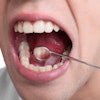
Cone-beam CT has the potential to become a new clinical tool for predicting mandibular invasion or erosion in patients with oral squamous cell carcinoma, according to a new study in the International Journal of Oral and Maxillofacial Surgery (March 6, 2010).
Researchers from Radboud University Nijmegen Medical Centre compared cone-beam CT, conventional preoperative panoramic radiography, MRI, and histological examination for assessing mandibular invasion of oral squamous cell carcinoma.
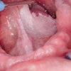
Between September 2006 and September 2009, the researchers assessed 23 patients with histology proven primary oral squamous cell carcinoma, adjacent to or fixed to the mandible. Their tumors were classified into four groups, ranging from no bone invasion to evident bone invasion.
Sensitivity and specificity for panoramic radiography were 55% and 92%, respectively -- both lower than the 91% sensitivity and 100% specificity for cone-beam CT, the researchers found. Comparatively, MRI showed 82% sensitivity and 67% specificity.
Although cone-beam CT was superior to both of these imaging methods in terms of sensitivity and specificity for this application, "its value may be limited by low sensitivity," the researchers noted. They plan to conduct a follow-up prospective study of 64 patients "to improve these results statistically," they concluded.
Copyright © 2010 DrBicuspid.com