While white-light examination (WLE) remains firmly entrenched as the most common screening method for oral and oropharyngeal cancers, brush biopsies and fluorescent/luminescent techniques are playing an increasingly important role in early detection of these diseases (Head & Neck Oncology, January 2009, Vol. 1:5).
“We picked up high-grade lesions that were not diagnosed under WLE, and that included dysplasias and carcinomas.”
— Mary Reid, Ph.D., Roswell Park
Cancer Institute
A study in this month's Cancer Prevention Research further validates the use of advanced imaging modalities -- in this case, autofluorescence-guided visualization (AFV) -- to enhance the detection of oral cancers, especially in high-risk patients (Cancer Prevention Research, November 2009, Vol. 11:2, pp. 966-974).
It also points to a gap in the adoption of these technologies by specialists and the need for the dental community to be more organized when it comes to diagnosing suspicious lesions, according to co-author Mary Reid, Ph.D., a research scientist in the Division of Cancer Prevention and Population Sciences at Roswell Park Cancer Institute.
Sequential surveillance
The Roswell study involved 60 patients who were considered high risk for oral cancer, because they had at least one of three criteria: presence of clinically suspicious oral lesions, a history of previously treated oral cancers with not evidence of recurrence for at least six month following treatment, or presence of recently diagnosed, untreated oral premalignant lesions or oral cancers.
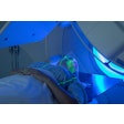
In addition to a conventional white-light exam, the patients underwent an AFV exam using a fluorescence imaging and point spectroscopy prototype that featured a 300-W xenon arc lamp illumination source operating at a wavelength of 405 nm (blue). (They used a prototype rather than a commercial AFV product because they were able to modify it with a cannula and go down into the throat, Reid explained.) A three-chip CCD video camera recorded the resulting images, and videos of both the white-light and AFV exams were stored on an attached computer.
Following a clinical examination and general oral hygiene assessment, detailed WLE and AFV exams of the oral cavity and parts of the oropharynx were conducted. Biopsies were then obtained from all areas found to be suspicious (n = 189), plus one normal-looking control area per person (n = 60). Because the study was intended to evaluate whether the addition of AFV to WLE improved the ability to detect cancerous lesions, sensitivity, specificity, and predictive values were calculated for WLE, AFV, and WLE plus AFV.
"Sequential surveillance with WLE + AFV provided a greater sensitivity than WLE in detecting low-grade lesions (75% vs. 44%), high-grade lesions (100% vs. 71%), and oral cancers (100% vs. 80%)," Reid and her colleagues wrote. In addition, 13 of the 76 additional biopsies obtained based on AFV findings were high-grade lesions or oral cancers, and five patients were diagnosed with a high-grade lesion or oral cancer only because of the addition of AFV to WLE.
However, the researchers noted, the specificity in detecting lesions decreased from 70% with WLE to 38% with WLE plus AFV. Even so, "AFV + WLE can be a highly sensitive first-line surveillance tool for detecting oral premalignant lesions and oral cancers in high-risk patients," they concluded. "Further refinement in autofluorescence technology and the development of adjunct genetic and molecular markers may be needed to improve the specificity of this technique."
Targeting oral surgeons
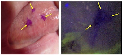 |
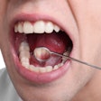
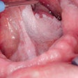
| In a study at Roswell Park Cancer Institute, 60 high-risk patients underwent sequential surveillance with white-light examination (left) and autofluorescence visualization (right) to determine if combining the two techniques improves early detection of oral cancers. Here, the autofluorescence image indicates a moderate dysplasia on the tongue. |
"If I were a dentist and I saw that AFV picked up cancers that I couldn't see under white light in people who have a lesion or a cancer, I would say that is important, totally worth it," she said. "In our study we picked up high-grade lesions that were not diagnosed under WLE, and that included dysplasias and carcinomas."
The study also illuminates the need for the dental community to have a common strategy when it comes to diagnosing and treating these patients, Reid emphasized.
"Dentists all over the country are very concerned about missing cancers, so they use tools such as the VELscope to detect lesions they can't see otherwise," she said. "But they don't biopsy -- they refer these patients out to oral surgeons who aren't screening with the VELscope [or other AFV equipment]."
This means that the oral surgeon may not see what the general dentist saw under fluorescent light and thus choose not to biopsy, she added.
"General dentists and oral surgeons need to be on the same playing field when it comes to the use of advanced modalities for oral cancer detection," said Martin Jablow, D.M.D. "It is a problem when a patient is referred to an oral surgeon (after using an advanced technique such as a VELscope, Identafi 3000, Vizilite, or Microlux Illuminator), and the surgeon dismisses the need to biopsy the area. This requires the dentist to either refer to another oral surgeon or continue to monitor an area that the patient now feels is okay because the specialist said so."
Dave Morgan, chief science officer at LED Dental -- the company that manufactures the VELscope -- said that many oral surgeons feel they don't need the technology because they are already good enough to do the diagnosis without it.
"Sometimes we labor under the misconception that things we see under AFV are not possible to see with WLE," he said. "But the majority of the time it is just subtle lesions that the general dentist would have otherwise missed. In fact, a lot of the time with the subtle lesions, if they see it with the VELscope first and then take the VELscope away, they will say, 'Oh, right, I see it now.' "
Photographic evidence
Once the lesion is referred to the oral surgeon for biopsy, having the VELscope can help, "but the key event has already happened: The lesion has been referred and you have an expert pair of eyes looking at it," Morgan added.
The company also recommends the primary practitioner include a white-light and VELscope image of the suspicious area with any referral.
"Sending pictures is always a good option to contextualize the case for the oral surgeon," Morgan said.
Still, he admits, "if we had our way, all oral surgeons would have these devices too." The company is actively marketing its products to oral surgeons, having most recently exhibited at the annual meeting of the Association of Oral and Maxillofacial Surgeons.
"We have many incentive programs in place to try and get oral surgeons to adopt this technology," he said.
