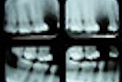
Orthopantomograms (OPTs) are not as effective as cone-beam CT images in evaluating miniscrew implant position prior to surgical intervention, according to a study in the Journal of Orofacial Orthopedics.
Researchers from examined the extent to which the OPT allowed miniscrew position to be estimated, and whether probands from different backgrounds in dental medicine arrived at the same conclusions regarding the screw position using the same images (Journ Oro Orth, May 2012, Vol. 73:3, pp. 236-248).
They inserted a tomas pin (8 mm x 1.2 mm) into all four quadrants of nine macerated skulls and then imaged the skulls with both OPT and cone-beam CT. The OPTs were then presented to orthodontists, oral and maxillofacial surgeons, and dental students who were asked to estimate the miniscrew position in relation to the neighboring structures. The skulls were also imaged using the Galileos cone-beam CT system (Sirona), and the results compared to the OPT evaluations.
None of the three groups estimated the screw's position in the OPTs significantly better or worse in comparison to the cone-beam CT images, the study authors found. However, their assessments of the miniscrew's proximity to the dental root differed and in many cases did not correspond exactly with the cone-beam CT measurement. In addition, the OPT assessments the proximity of the screw to the maxillary sinus and/or inferior alveolar nerve differed from the proximity measured in the cone-beam CT, according to the researchers.
The study findings demonstrate that OPTs can achieve a rough evaluation of the miniscrew position in relation to the surrounding structures. However, the authors noted, practitioner assessments were "imprecise," with "major deviations" in their determination of the screw's proximity to the dental root.
"For this reason, if there is any doubt, patients should take advantage of three-dimensional diagnostics, preferably before any surgical intervention," the researchers concluded.


