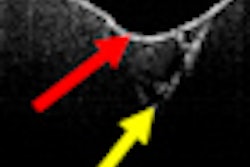
Optical coherence tomography (OCT) is an efficient, noninvasive method for detecting the depths of lesions caused by demineralization, according a study in Brazilian Oral Research (September/October 2011, Vol. 25:5, pp. 407-413).
In addition to its ability to identify caries and monitor caries progression without the use of ionizing radiation, OCT has the potential to determine the locations and depths of lesions, and detect hidden and secondary caries that occur at the tooth-restoration interface, according to the study authors, from the University of São Paulo.
For these reasons, they chose OCT to evaluate the degree of demineralization that would occur in artificially induced caries-affected human dentin.
First, the researchers abraded the occlusal surfaces of six human molar teeth until a flat surface was obtained and the enamel was removed to expose the occlusal dentin surface. They then sectioned the teeth into 12 halves in the vestibular-lingual direction and divided them into three groups according to the period length of their exposure to a microbiological assay (n = 4): G1, 7 days; G2, 14 days; and G3, 21 days.
The surfaces of all specimens were protected by an acid-resistant nail varnish, except for a window where the caries lesion was induced by a Streptoccocus mutans biofilm in a batch-culture model supplemented with 5% sucrose.
The researchers then analyzed the specimens with OCT and obtained qualitative and quantitative results (images and average dentin demineralization, respectively). The mean demineralization depths (in micrometers) obtained using OCT are listed below:
- Group 1: 235 ± 31.4 µm
- Group 2: 279 ± 14 µm
- Group 3: 271 ± 8.3 µm
"The images obtained by OCT confirm that this method is effective in evaluating the depth of demineralization caused by induction of caries in vitro from 7 days on," the researchers wrote. "The images clearly show that the demineralization process is occurring in the specimens after different periods of exposure to the bacterial challenge."


