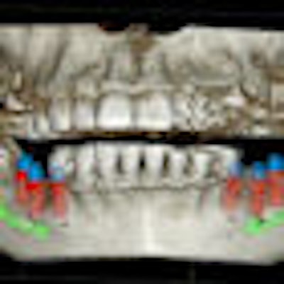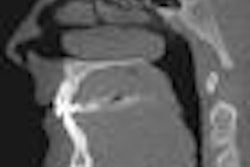
With the advent of more affordable cone-beam CT systems (CBCT), a growing number of dental practitioners are investing in this equipment. But the increasing adoption of CBCT begs a parallel demand for users to correctly analyze and diagnose the resulting 3D volume datasets.
One inherent aspect of CBCT that makes this challenging is the presence of artifacts, such as noise, scatter, motion missing values, beam hardening, and rings. These artifacts are often the result of discrepancies between the physical imaging process and mathematical modeling in the reconstruction algorithms, according to researchers from Johannes Gutenberg University and University of Applied Sciences in Germany (Dentomaxillofacial Radiology, July 2011, Vol. 40:5, pp. 265-273).
To understand what causes CBCT artifacts and how they can impact diagnostic accuracy, it is useful to understand the underlying reconstruction principle in CBCT and CT: backprojection. From a technical point of view, all CBCT machines reconstruct the volume from a high number of two-dimensional x-ray projections acquired in a circular orbit around the target, the researchers noted. However, "in the technical community it is well known that reconstructions from circular orbits are insufficient for an accurate reconstruction of the volume," they wrote.
“Artifacts are inherent to the design of the device.”
— Ed Marandola, Imaging Sciences
International
In addition, most CBCT systems use the Feldkamp reconstruction algorithm, which applies "a simple approximate weight to the projection values instead of using the actual analytically computed distances that the measured 'rays' have traveled from source to detector," the study authors noted. "All of these factors, in combination with other insufficiencies inherent in the measurement and reconstruction process, introduce artifacts into the cone-beam datasets."
Since these artifacts may interfere with the diagnostic process, "every user should be aware of their presence," they wrote. However, "there seems to be a knowledge transfer gap between the technical and the radiographic community ... [that] may introduce diagnostic errors that could be avoided by a better understanding of the causative factors and error factors."
Typical artifacts
An artifact is "a visualized structure in the reconstructed data that is not present in the object being imaged," according to the German researchers. Artifacts most often represent as streaks, line structures, and shadows along the projection lines, they noted.
"Artifacts are inherent to the design of the device, and the design of the device is always a set of compromises," said Ed Marandola, a founder and former president of Imaging Sciences International (ISI), which manufactures the i-CAT CBCT system. "But they are true of any imaging method. There is always some kind of artifact and something being done behind the scenes to remove it."
The following are some common artifacts:
Noise: Noise is considered one of the most common artifacts in CBCT imaging. There are two types of noise in reconstructed CBCT images: additive, stemming from round-off errors or electrical noise, and photon count. Noise represents itself in inconsistent attenuation values in the projection images -- a "graining" on the image.
"Noise will always be one of the big issues," said Marandola, who is currently senior vice president of technology and innovation for Gendex, Dexis, and ISI. "But it is always improving. The detector we use today is very different from what we used when we first introduced the i-CAT in 2004-2005."
Scatter: In radiographic imaging, only photons traveling directly from the source to the detector are measured, according to the study authors. Scatter is caused by those photons that are diffracted from their original path after interacting with the object being imaged. The larger the detector, the higher the probability that scattered photons incite it, the researchers noted.
"Scatter is the really big one, and this is where the big difference between CBCT and medical CT occurs," Marandola said. "In a CBCT image, the whole area you are looking at is all of the image, so when you get scatter, it scatters all through the image. In medical CT, the scatter is significantly less because the detector is very small."
Partial volume effect: This appears as "blurring" over sharp edges and is due to the scanner being unable to differentiate between a small amount of high-density material and a larger amount of lower density material. When the processor tries to average out the two densities or structures, information is lost.
Beam hardening: This occurs when there is more attenuation in the center of the object than around the edge. The result is often streaking or a "cupped" appearance.
"Beam-hardening artifacts are why the i-CAT operates at 120 kpv and is heavily filtered," Marandola said. "We heavily filter the x-ray beforehand so that we have such a hard beam that it can penetrate the skull, rather than relying on the skull to filter the beam. It is expensive to do, but it helps to eliminate the beam-hardening artifacts."
Ring artifacts: Ring artifacts are one of the most common mechanical artifacts and are often the result of a detector default. They appear as concentric rings centered around the axis of rotation. Using a smaller flat panel with the source-detector axis positioned offset to the center of rotation can result in ring artifacts, according to the study authors.
"The most expensive component on a CBCT is the sensor, and the larger the sensor the more expensive it is," Marandola said. "But the smaller the panels get, the more problems you run into. Consider the approximation techniques in backplane reconstruction -- the entire volume you are looking at is in the whole image. Say you are scanning a soup can. When the object is inside the can, it is harder to know where the x-ray started. That is why the smaller volume machines run into problems. You are looking inside the skull, but you didn't start from outside the skull."
Motion/misalignment: This is when blurring and/or streaking occurs due to movement by the object being imaged.
"There are head holders to keep patients from moving, but motion artifacts are probably the single biggest thing that doctors see in a CBCT image, and they probably don't even realize it when they see them," Marandola said. "If we want to progress with CBCT to more customized applications such as custom implants or even Invisalign, that's when motion becomes very important. The company that comes out with motion correction is going to be able to drive CBCT into the next level for the dentist."
Correcting for artifacts
The study authors also point to the inherent "general inconsistencies in reconstructions" found in small field-of-view (FOV) CBCT, noting that "in small field-of-views ... the FOV is just a small part of the entire object that is being traversed by the x-rays." There are structures outside of the FOV that are only in the beam over small angular ranges, they added, "yet the backprojection process does not account for that. There is no easy way to overcome this problem, which again causes inconsistencies (errors) in the reconstructed volume."
Marandola agreed and maintains that large FOV systems offer many advantages for this reason.
"The trend to small FOV has been driven by cost and radiation dose," he said. "Cost is cost, and you can't ignore that -- some doctors just can't afford this technology, and in some cases small FOV is all they need. But I tend to differ with the radiation issue in most cases. You can always control the height, and radiation dosages are not as severe as people tend to make them sound."
So what does all this mean for dentists using CBCT in daily practice?
"It is no surprise that the technical community puts considerable efforts into developing techniques for artifact reduction," the study authors wrote. "Many of them are postprocessing algorithms operating on the 3D volume data. Although this may result in considerable reduction of some apparent artifact structures, from a physical point of view postprocessing is like putting the cart before the horse since the error has been integrated into the volume already."
One recent solution to this problem is to avoid reconstruction errors altogether by supplementing missing or incorrect information in the projection images or by integrating some sort of "meta-information" into an iterative reconstruction process, they added.
"There is software and all kinds of things you can do to an image both prior to reconstruction and after," Marandola said. "But you can't change the characteristics of the receptor panel itself. It's like taking a picture of a person -- the prettier the person, likely the better the picture will be. The photograph can affect lighting and even touch things up after the fact. So you can do a certain amount [to improve the image], but only so much."
Practitioners should have knowledge of the types of artifacts that appear on CBCT scans and how to best avoid them through resolution choices and scan options, he added.
“Depending on the type, size and location of the artifact, it could conceivably negatively affect diagnosis, but we feel these instances would be rare if doctors are using good imaging protocols,” he said. “If the dentist sees something that is unexpected or inconsistent with the anatomy being x-rayed, an oral radiologist can be consulted.”
Enhanced reconstruction methods will likely be more common in the near future, which will help to reduce many artifacts, the study authors concluded.


