Dear Imaging & CAD/CAM Insider,
Lots of cone-beam CT news and research in this Imaging & CAD/CAM Insider. First up: Orthodontists may have been some of the earliest adopters of cone-beam CT in the dental field, but did some practitioners jump the gun?
With the majority of orthodontic patients being young teens who often undergo full-head imaging before, during, and after treatment, there is concern that using cone-beam CT in every case unnecessarily exposes these patients to dangerous amounts of radiation.
Click here to read our latest Imaging & CAD/CAM Insider Exclusive, including what one imaging expert says practitioners need to know in order to minimize dose and maximize the quality of cone-beam CT imagery in orthodontics.
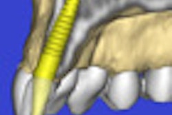
In other cone-beam CT news featured in the Imaging & CAD/CAM Community, this technology's ability to create 3D images that contain key anatomical and morphological details is also expanding its use in implant treatment planning. In fact, some experts say it is quickly becoming the standard of care in implantology. Read more.
And an alignment device created by British researchers may help dental practitioners better position each patient undergoing cone-beam CT imaging and eliminate the need for preliminary scout views prior to scanning. Click here to learn more.
In CAD/CAM news, have you been looking for an easier, less costly way to create dental bars? An invention by researchers from Missouri University of Science and Technology that entirely digitizes and automates the process could be the answer. Read more.
And a number of new CAD/CAM products have hit the market in recent months, including new scanners, systems, software, and design tools.
Finally, post-treatment endodontic images can significantly enhance post-mortem forensic identification, according to a report in the Australian Endodontic Journal. The key is in the quality of the ante-mortem radiographs obtained by general dentists and made available to forensic scientists.


