Clinician confidence is higher for cone-beam CT than for conventional radiographs in the diagnosis and treatment planning of maxillary impacted canines, according to a study in the American Journal of Orthodontics and Dentofacial Orthopedics (May 2010, Vol. 137:5, pp. 590-597).
Researchers from the University of California, San Francisco identified 25 consecutive impacted maxillary canines from a pool of patients seeking orthodontic treatment and compared them using two imaging modalities.
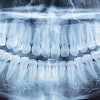
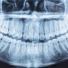
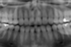
The first set of radiographs consisted of traditional 2D images, including panoramic, occlusal, and two periapical radiographs. The second set comprised prints of 3D volumetric dentition images obtained from a cone-beam CT scan. Seven faculty members completed a questionnaire for every impacted canine and diagnostic modality.
The judges produced different decisions regarding localization depending on the x-ray method, the researchers found. There was 21% disagreement in the perceived mesiodistal cusp tip position and 16% difference in the perceived labiopalatal position. In the perception of root resorption of adjacent teeth, there was 36% lack of congruence.
Twenty-seven percent of teeth that were planned to be left, recovered, or extracted with the radiographs had different treatment plans when the judges viewed the cone-beam CT images (p = 0.035). The clinicians' confidence of the accuracy of the diagnosis and treatment plan was statistically higher for cone-beam CT images (p < 0.001).
According to these findings, the researchers concluded that 2D and 3D images of impacted maxillary canines can produce different diagnoses and treatment plans.
Copyright © 2010 DrBicuspid.com