An accidentally swallowed 2-cm piece of an orthodontic aligner, which was difficult to detect because it was clear, was retrieved from a patient after imaging window adjustments were made. The case report was published on September 17 in BMC Oral Health.
Clinicians should warn patients not to wear fractured orthodontic appliances with low retention force until they discuss it with their treating dentists. This report is believed to be the first case of a patient swallowing part of their invisible aligner, the authors wrote.
"For invisible braces, impression materials, resins and other materials with low X-ray opacity, CT (computed tomography) should be taken and the lung window should be selected for image reading," wrote the authors, led by Dr. Yichen Pan of the Shanghai Ninth People's Hospital in China.
A 31-year-old female with poor occlusion
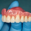
To address the patient's open bite and crowding, clinicians designed intrusion of maxillary first and second molars by mini-screws, with maxillary expansion by aligners. Clear aligners were used to help maintain the width of the woman's upper molars.
About three months later, her aligner cracked between her upper right second premolar and first molar. Despite the fracture, she continued to wear the aligner without notifying her clinician. While she was drinking water, the woman accidentally swallowed a piece of the aligner, the authors wrote.
She felt pain while swallowing so she went to an emergency room. A laryngoscopy revealed no foreign body. Since the plastic of the aligner makes it difficult to see clearly on x-rays, she underwent a computed tomography (CT) scan. Still, no foreign object could be seen, so the radiology department was consulted.
The grayscale range to the lung window of the CT was adjusted, and clinicians saw a strip-shaped foreign body in the woman's esophagus. It was decided that the broken aligner would be removed via endoscopic surgery, they wrote.
 CT images of the esophageal foreign body. (A) Transverse section on the T2 level in the soft-tissue window. (B) Transverse section on the T2 level in the lung window. (C) Sagittal section of the esophagus and the foreign body in the lung window. Images and captions courtesy of Y. Pan, J. Huang, Y. Xie. Licensed under CC BY 4.0.
After superficial anesthesia was administered, forceps were used to remove the fractured aligner from the woman's esophagus. After three days, the pain she felt while swallowing was gone. At her one-month follow-up appointment, the woman had no adverse symptoms, the authors wrote.
 Endoscopic images. (A, B) Image of the foreign body in the woman's upper esophagus. (C) Foreign body taken out with a clamp. (D) The foreign body in vitro.
Unique situation
When a foreign body is lodged in the esophagus, it needs to be retrieved because it's nearly impossible to expel itself. Imaging must be used to confirm the location and size of a foreign body.
 Fractured pieces of the orthodontic invisible aligner. (A) The 2-cm swallowed piece of the aligner. (B) Fractured pieces of the whole aligner on the 3D-printed model. (C) Fractured pieces of the whole aligner.
However, this case was unusual because typically swallowed dental instruments often contain high-density components such as metal and ceramic, while orthodontic invisible aligners are made of clear plastic, which has low x-ray opacity. Therefore, adjustments to the gray value range to the lung window needed to be necessary to best visualize the object.
Accidental ingestion or aspiration of a dental appliance can cause damage or threaten a person's life, the authors wrote.
"To avoid accidental ingestion, patients need to be told that when poor retention, crack or fracture occurs in any appliance, they should contact the attending doctor immediately and do not continue to wear it," Pan et al wrote.
