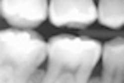
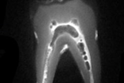
Certain near-infrared (NIR) imaging wavelengths offer better contrast for imaging tooth demineralization than others, according to a new study in Lasers in Surgery and Medicine (July 16, 2013).
In vivo and in vitro studies have shown that high-contrast images of tooth demineralization can be acquired in NIR without the interference of stain, noted the study authors, from the University of California, San Francisco.
They painted 24 human molars with an acid-resistant varnish, leaving a 4 x 4-mm window in the occlusal surface of each tooth exposed for demineralization. Artificial lesions were produced in the exposed windows after one- and two-day exposure to a demineralizing solution. The lesions were imaged using NIR reflectance at three wavelengths: 1,300, 1,460, and 1,600 nm. They were also imaged with visible light reflectance and fluorescence for comparison.
The contrast of the one- and two-day lesions were significantly higher (p < 0.05) for NIR reflectance imaging at 1,460 and 1,600 nm than it was for NIR reflectance imaging at 1,300 nm, visible reflectance imaging, and fluorescence, the researchers found.
These findings suggest that the 1,460- and 1,600-nm wavelengths are better-suited than 1,300 nm for imaging early/shallow demineralization on tooth surfaces, they concluded.


