Magnetic resonance imaging (MRI) offers several advantages over conventional radiography when diagnosing dental abnormalities in children and for orthodontic treatment and surgical planning, according to a new study in the Journal of Oral and Maxillofacial Surgery (April 20, 2013).
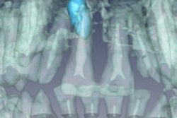
Researchers from the University of Würzburg selected 16 patients (mean age, 10.8 years) from 1,500 orthodontic patients: three with a mesiodens, nine with supernumerary teeth other than a mesiodens, one with gemination, one with dilacerations, one with transmigration, and one with transposition. Three-dimensional images were acquired on a 1.5-tesla MRI system at measurement times of 4 to 5 minutes.
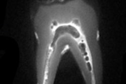
Using natural MRI contrast, the researchers were able to clearly delineate the teeth, dental pulp, mandibular canal, and cortical bone of each patient. In addition, the position and shape of malformed teeth could be assessed in all three spatial dimensions, they noted.
"MRI was found to be a well-tolerated imaging modality for the diagnosis of dental abnormalities in children and for orthodontic treatment and surgical planning," the study authors concluded. "Compared with conventional radiography, dental MRI provides the advantage of three-dimensionality and complete elimination of ionizing radiation, which is particularly relevant for repeated examinations in children."