Cone-beam CT (CBCT) offers advantages over periapical radiographs for treatment planning of mandibular molars undergoing apical surgery, according to a new study in the Journal of Endodontics (February 2011, Vol. 37:2, pp. 151-157).
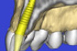
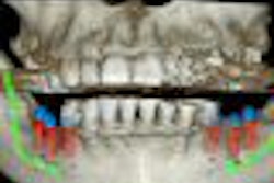
Researchers from the University of Bern compared cone-beam CT to conventional periapical radiographs in evaluating 38 mandibular molars with 75 roots prior to apical surgery. They looked at the detectability and dimensions of periapical lesions, the relationship of the mandibular canal to the roots of the respective teeth, and the dimension of the buccal bone.
Of 58 detected periapical lesions, 15 (25.9%) lesions diagnosed with sagittal cone-beam CT slices were missed with periapical radiography, the researchers found. The distance between the apices and the upper border of the mandibular canal was only measurable in 24 of 68 radiographs (35.3%) by using periapical images. The cortical bone wall had a mean thickness of 1.7 mm, whereas the total buccal bone wall measured 5.3 mm on average.
"The present study highlights the advantages of using limited CBCT for treatment planning in mandibular molars before apical surgery," the researchers concluded.
Copyright © 2011 DrBicuspid.com