Dear Imaging & CAD/CAM Insider,
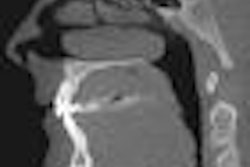
The growing use of cone-beam CT for diagnostic and treatment planning applications in dentistry has prompted a parallel body of discourse on the legal liabilities associated with 3D image interpretation.
These discussions have, in turn, prompted a number of surveys of incidental findings in dental cone beam CT images, including two presentations at the recent American Academy of Oral and Maxillofacial Radiology (AAOMR) meeting. Read more about the potential legal ramifications of incidental findings in this latest Imaging & CAD/CAM Insider Exclusive.
In other news from the AAOMR meeting:
- An interactive software tool being developed at the Creighton University School of Dentistry automates the reading of dental CT scans by recognizing and labeling key anatomical features, and its developers think it could speed up the training required to interpret these complex 3D images.
- When more than half of his sophomore class indicated they use an iPhone on a daily basis, a radiology professor at Case Western Reserve University developed two iPhone/iPad apps designed to make it easier -- and more fun -- for the students to learn the anatomy they encounter in panoramic radiographs and cone-beam CT images.
Meanwhile, dental cone-beam CT made the front page of the New York Times last month in an investigative report that painted a less-than-flattering picture of the technology's proponents. But imaging experts and those who design and sell the equipment say the Times missed some key facts, and that when it comes to reducing radiation exposure, users bear some responsibility as well. Read more by clicking here or visit the Imaging & CAD/CAM Community.
The issue is already being addressed by some government agencies and industry organizations. On November 8, the U.K. Health Protection Agency released new guidance on the use of cone-beam CT in dentistry; that same day, the AAOMR and the American Association of Endodontists issued a joint position statement on the use of cone-beam CT in endodontics.
These concerns could open the door for new diagnostic tools that rely on optical sources rather than ionizing radiation. Researchers from the University of Rochester are using Raman spectroscopy -- a highly sensitive microscopy technique -- to study two common dental plaque bacteria, with an eye toward developing a new oral health assessment and caries detection tool. Read more.