There is a need to better understand radiographic features associated with bisphosphonate exposure and to determine whether osteosclerosis is a specific finding indicative of the risk for progression to bisphosphonate-related osteonecrosis of the jaw (BRONJ) according to a study in the Journal of Oral and Maxillofacial Surgery (September 2010, Vol. 68:9, pp. 2232-2240).
"Radiographic features in patients with bisphosphonate-related osteonecrosis of the jaw (BRONJ) are well described, but less is known in bisphosphonate-exposed individuals with stage 0 disease (clinical symptoms without exposed necrotic bone) considered at risk for BRONJ," wrote researchers from Kaiser Permanente Oakland Medical Center and Highland General Hospital in California. Their goal with the study was to identify imaging features that may presage development of BRONJ.
A dental symptom survey was returned by 8,572 Kaiser Permanente health plan members receiving chronic oral bisphosphonate therapy, and the researchers examined 1,005 patients who reported pertinent dental symptoms or complications after dental procedures. Those without BRONJ but with concerning symptoms were referred for clinical evaluation, including imaging. Among the subset who received maxillofacial imaging, the researchers identified those with stage 0 disease and abnormal radiographic features.
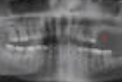
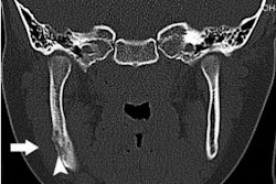
They found a total of 30 patients without exposed bone but with concerning symptoms who received maxillofacial imaging (panoramic radiography or computed tomography) in the context of clinical care. Among these 30 patients, 10 had stage 0 disease with similar radiographic features of regional or diffuse osteosclerosis in clinically symptomatic areas, most with extension beyond the involved site.
Other findings in these 10 patients included:
- Density confluence of cortical and cancellous bone
- Prominence of the inferior alveolar nerve canal
- Markedly thickened and sclerotic lamina dura
- Uniform periradicular radiolucencies
- Cortical disruption
- Lack of bone fill after extraction
- A persisting alveolar socket
None of the 10 patients had exposed bone develop during one year of follow-up. The remaining 20 patients had normal or localized radiographic findings consistent with odontogenic pathology, the researchers noted.
Copyright © 2010 DrBicuspid.com