Cone-beam CT (CBCT) can better identify the location of inferior alveolar nerves (IANs) in relation to impacted mandibular third molars during preoperative evaluations, according to research presented in June at the Computer Assisted Radiology and Surgery conference in Geneva.
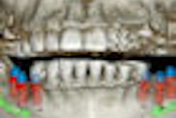
An uncommon but serious complication of mandibular third-molar removal is injury to the IAN, according to Vicente Hernandez Soler, M.D., D.D.S., M.Sc., of Valencia University in Spain. Panoramic radiographs have long been used to assess third-molar eruption, but they do not show the buccolingual position of the canal, he noted. In comparison, cone-beam CT has the advantage of high resolution and low dose and can identify cortication of the mandibular canal.
Comparing panoramic x-rays and cone-beam CT images, Dr. Soler and his colleagues examined the buccolingual relationship between mandibular third molars and the mandibular canal and assessed which view -- panoramic, axial, or cross-sectional -- provided better visibility for mandibular canal identification at different levels.
They evaluated 20 patients who had had orthodontic treatment and were at risk of third-molar impaction, based upon review of post-treatment panoramic radiographs. An i-CAT cone-beam CT system (Imaging Sciences International) was used to assess the IAN/third-molar relationship.
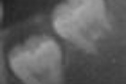
"The results showed that cone-beam CT cross-sectional images as well as reconstructed panoramic images gave a clear perception of the mandibular nerve," Dr. Soler noted. Cross-sectional is the best view anterior to the third molar, while panoramic is the best view posterior to the third molar, he added.
"Advanced 3D reconstruction to evaluate third molars provides information beyond that provided by panoramic images" and confirmed the clinical usefulness of cone-beam CT for preoperative evaluation of impacted mandibular third molars, he concluded.
Copyright © 2010 DrBicuspid.com