Dear Imaging & CAD/CAM Insider,
Nearly every laser in dentistry is considered by the U.S. Food and Drug Administration to be a class IV device, meaning they pose potentially serious eye, skin, fire, diffuse reflection, and plume radiation risks.
These same risks apply whether you use an erbium hard-tissue laser, an Nd:YAG laser for periodontal surgery, or one of the new handheld diode lasers coming onto the market. In fact, with their greater mobility, these compact, portable devices -- which look much like laser pointers on steroids -- could pose new challenges for a dental practice's laser safety officer.
Read more about how to ensure you and your staff are adequately trained and using the right safety equipment in our latest Imaging & CAD/CAM Insider Exclusive.
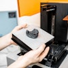
In other imaging news, a Canadian company is gearing up to launch a novel caries detection device later this year and will present some of its first clinical results at the International Association for Dental Research (IADR) meeting in July. Click here to read what makes this product different from those already on the market.
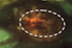
Optical technology is also at the heart of a portable imaging system for oral cancer screening developed at Rice University to improve cancer survival rates in developing countries as well as industrialized nations. Findings from a recent clinical study in India show the device demonstrates both high specificity and high sensitivity. Read more.
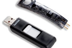
And will intraoral cameras one day make their way into patients' homes, allowing them to practice "personal diagnostics"? That's the dream of one company that is currently testing its virtual diagnosing and networking system. Read more.
In research news, a study out of the University of Bonn found that dental practitioners can aid in the early detection of strokes by using panoramic radiographs to identify radiopaque findings in the carotid region, while researchers from Malmo University found no clear evidence that patients suspected of temporomandibular disorders should be examined using any imaging, according to a study in the Journal of Oral Rehabilitation.
Finally, in other research to be presented at the IADR meeting, cone-beam CT scans were found to be "significantly more efficient" than periapical radiographs in determining the mesiobuccal root canal anatomy of maxillary molars prior to endodontic treatment. Read more by clicking here.