Cone-beam CT can improve the detection of proximal-surface caries extending into the dentin but cannot do the same for occlusal caries, according to researchers from the University of California, Los Angeles School of Dentistry (Dentomaxillofacial Radiology, October 2009, Vol. 38:7, pp. 445-451).
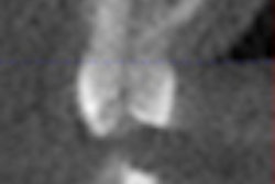
They compared cone-beam CT images produced by a 3DX Accuitomo (J. Morita) to conventional charge-coupled device (CCD) images in nonrestored, extracted human permanent premolar and molar teeth.
They selected 92 occlusal and 100 proximal surfaces; 36 of the occlusal and 25 of the proximal had lesions extending into dentin. Using a five-step confidence scale, eight practicing dentists evaluated the images for the presence of caries in dentin using both modalities. Actual presence and extent of caries was established with microCT imaging.
For proximal surface lesions extending into dentin, the average sensitivity score using 3DX images (0.61) was almost twice that of CCD images (0.33), and the difference was significant, the researchers reported. The specificity values for both systems were high and not significantly different from each other.
For occlusal surfaces, the raters detected significantly more lesions in the enamel or dentin when using the 3DX images than when using CCD images. However, the raters also had significantly lower average specificity scores for the 3DX images compared with the CCD images for lesions at both depths, the researchers reported.
Copyright © 2009 DrBicuspid.com