Case presentation
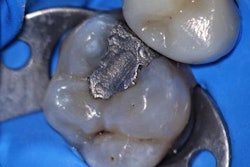
A 40-year-old man presented with sensitivity in tooth #4 and discomfort during mastication. Clinical examination revealed caries on the distal wall of tooth #4, extending close to the pulp chamber.
 Dr. Jorge F. Zapata.
The patient desired a metal-free, aesthetic restoration and relief from sensitivity. The treatment plan involved a Class II pulp cap restoration using immediate dentin sealing (IDS) and resin coating technique (RCT) followed by composite resin placement for a functional and aesthetic restoration.
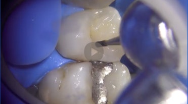 Preop screenshot of tooth #4. Images courtesy of Dr. Jorge F. Zapata.
Treatment plan
The primary goal of the treatment was to protect the exposed dentin and pulp, eliminate sensitivity, and restore function and aesthetics to the tooth using an advanced composite resin. The chosen materials were Clearfil SE Protect adhesive and Clearfil Majesty ES-2 Classic, both from Kuraray Noritake Dental, selected for their superior handling, aesthetic integration, and durability. Watch the full clinical procedure.
Clinical procedure
- Anesthesia and isolation: Local anesthesia was administered to ensure patient comfort throughout the procedure. Tooth #4 was isolated using a rubber dam to maintain a clean, dry operative field and prevent contamination from saliva and moisture.
- Removal of carious tissue: Using a high-speed handpiece with carbide burs, the decayed dentin was carefully removed. Special attention was given to preserving as much healthy tooth structure as possible, particularly around the pulp chamber, to avoid further trauma. Caries Detector (Kuraray Noritake Dental) was applied to ensure all compromised dentin was removed prior to pulp capping.
- Immediate dentin sealing: To protect the dentin, a primer (Clearfil SE Protect) was applied for 20 seconds followed by bonding for 10 seconds. The bonding agent was then light cured. This step, known as IDS, creates a protective seal over the dentin to prevent contamination and sensitivity.
- Pulp capping: Upon inspection, the decay had extended close to the pulp, but the pulp chamber itself remained intact. Limelite (Pulpdent) pulp liner was applied to the affected area to promote healing and provide a protective barrier over the pulp. A resin-modified glass ionomer (Vitrebond by 3M) was used to provide additional protection and support for bonding.
- Resin coating technique: A thin layer of flowable composite (Clearfil Majesty ES Flow by Kuraray Noritake Dental) was applied to the pulpal floor to adapt to the irregularities and create a smooth surface. This layer was light cured for 20 seconds.
- Composite resin placement
- Base layer: A flowable composite (Clearfil Majesty ES Flow) was applied to the pulpal floor, further enhancing adaptation to the cavity walls.
- Incremental buildup: A nanofilled composite resin in shade A2 (Clearfil Majesty ES-2 Classic) was placed in 2-mm increments to minimize polymerization shrinkage and ensure proper curing. Each increment was shaped and contoured to the natural anatomy before light curing for 20 seconds.
- Interproximal restoration: A sectional matrix system was used to restore the interproximal wall, ensuring proper contact and contour. The matrix band was maintained until the composite was fully polymerized to avoid deformation.
- Finishing and polishing: The occlusal anatomy was sculpted and refined using finishing burs. Additional light curing was performed to ensure complete polymerization. The restoration was polished using a Sof-Lex (3M) polishing strip for interproximal surfaces and an Enhance Finishing cup (Dentsply Sirona) for the occlusal surface, followed by Jiffy polishers (Ultradent) to achieve a high-gloss, enamel-like finish.
- Occlusion adjustment: The rubber dam was removed, and the patient's occlusion was checked using articulating paper. Minor adjustments were made to eliminate premature contacts, ensuring a comfortable and functional bite.
Outcome and follow-up
The procedure successfully restored tooth #4 with an aesthetic, functional, and durable composite restoration. The patient expressed satisfaction with the improved appearance and reported an immediate cessation of sensitivity. A follow-up examination confirmed the restoration's stability, proper occlusion, and the absence of any postoperative complications.
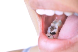
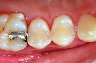 Postop photo of tooth #4.
Discussion
Pulp cap restorations require a meticulous technique to ensure pulp protection and long-term success. The IDS technique combined with the RCT offered multiple benefits, including improved bond strength and reduced sensitivity.
The use of an advanced composite resin provided several key advantages, including the following:
- Handling and manipulation: The composite allowed for precise placement and contouring without slumping or sticking to instruments.
- Aesthetic integration: The composite's light-diffusion properties achieved a seamless blend with the surrounding tooth structure, mimicking natural enamel.
- Physical properties: The material exhibited low polymerization shrinkage and high mechanical strength, contributing to the restoration's durability.
- Polishability: The composite achieved a natural-looking finish, enhancing aesthetics and helping to reduce plaque accumulation.
Conclusion
This case illustrates the effective use of IDS and RCT in a class II pulp cap restoration using a nanofilled composite resin. Following clinical protocols -- from dentin sealing and bonding to incremental composite placement and polishing -- resulted in a durable and aesthetically pleasing restoration. The patient's positive feedback and the restoration's functional integrity underscore the benefits of high-quality materials and adherence to best practices.
The comments and observations expressed herein do not necessarily reflect the opinions of DrBicuspid.com, nor should they be construed as an endorsement or admonishment of any particular idea, vendor, or organization.
Dr. Jorge F. Zapata is a general dentist in Ogden, UT, who has been practicing microscope dentistry in his private dental practice since 2005. He is a past executive board member and former treasurer of the Academy of Microscope Enhanced Dentistry. Additionally, he serves as a key opinion leader for Kuraray Noritake Dental and Pascal Dental. He can be contacted at [email protected].


