Clinicians used a new surgical technique to remove a separated endodontic instrument stuck about 2 mm below the tip of a patient's tooth root in a case published in the Journal of Endodontics.
They performed an intentional tooth replantation along with root resection under local anesthesia to pull the broken instrument from a Japanese woman's tooth extraction socket. Because the management of broken endodontic instruments is contentious, clinicians should consider all surgical and nonsurgical options when developing treatment plans, according to the authors.
"We planned a novel surgical approach, which reduces the damage to the tooth, bone, and nerves, to remove the extruded instrument at the patient's request," wrote the group, led by Dr. Takeshi Harada from the oral and maxillofacial surgery department at Osaka University Graduate School of Dentistry in Japan (J Endod, September 2, 2021).
Technique used to extract the instrument
In June 2019, a 24-year-old woman was referred to the hospital after a general dentist failed to remove a separated endodontic instrument with a nonsurgical approach. The dentist's failure to manage the broken instrument resulted in it extending beyond the root apex and migrating into her jaw, which the hospital confirmed with an x-ray and computed tomography (CT) scan. Additionally, CT showed that the instrument ran along the mandibular lingual cortical bone with a curve and touched the border of the intramandibular joint.
After talking with the woman, the emergency clinician decided to treat her with intentional tooth replantation along with alveolar osteotomy while the patient was under local anesthesia.
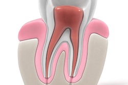
Prior to the replantation, the clinician prepared the tooth's root canal while continuously irrigating the root of the tooth with 3% sodium hypochlorite. The dentist flushed with 5 mL of distilled water, dried with paper points, and used a cone with a sealer to fill the canal. The canal was then compacted laterally with multiple accessory cones, and composite resin was used to seal the access cavity, the authors wrote.
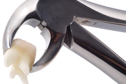
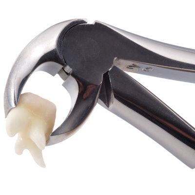
The clinician used forceps to pull the woman's second premolar. The dentist placed the forceps on the tooth's crown and used a slow rocking and rotating motion to prevent periodontal ligament damage. Until the second premolar could be replanted, it was kept in sterilized gauze filled with saline.
After the premolar extraction, the clinician could see the top end of the separated instrument at the bottom of the patient's tooth socket; however, removal was challenging, with a risk of breakage and pushing it in more so that it could no longer be seen.
A gingival sulcular incision was made, and the clinician performed a vertical osteotomy on the buccal aspect proximal and distal to the tooth socket and a horizontal osteotomy at the bottom of the socket. To maintain blood circulation, the osteotomy bone segment was attached to the mucoperiosteal flap for the duration of the procedure.
Then, the clinician performed piezoelectric bone surgery to remove bone marrow around the separated instrument. This allowed the clinician to extract the instrument buccally and upward without excessive force. A postoperative x-ray confirmed the instrument's complete removal, they wrote.
Successfully finishing the procedure
Because of the separated instrument, the original dentist had not been able to fill the root canal of the extracted tooth properly. The emergency clinician performed a root-end resection 2 mm from the apex of the patient's root canal and prepared a 1-mm root-end cavity. The root-end cavity was washed, air-dried, and filled. The clinician then restored the mucoperiosteal flap and osteotomy bone segment to their original positions.
Finally, the extracted premolar was replaced by hand. The surgeon exerted pressure in the axial direction and used sutures to better adapt the socket wall to the tooth root.
For four weeks, twisted stainless steel wire and composite resin were applied to stabilize the tooth. At follow-up visits at two, four, six, and 12 months, the patient reported no pain or other complications.
After a year, the tooth's periodontal ligament cavity showed no signs of inflammatory absorption. Also, a CT scan showed bone marrow regenerated around the tooth, and it revealed continuity of the buccal cortical bone, the authors noted.
New strategy, common problem
Despite innovations in their design and composition, endodontic instruments can separate within the root canal in patients. Separation occurs up to 5% of the time and can hinder cleaning and shaping during root canals, which could lead to endodontic treatment failure.
Clinicians can manage separated instruments with surgical and nonsurgical procedures, but there are no perfect solutions. Surgical procedures have success rates between 55% and 79%, and nonsurgical management can result in other complications, such as pushing the separated instrument deeper.
Though the new procedure was successful in this case, the authors noted that the use of intentional replantation is limited by periodontal ligament conditions and root forms.
Nevertheless, "it is important to consider all options, including surgical approaches, when establishing the treatment plan," Harada and colleagues wrote.


