Imaging has helped diagnose and reconstruct an 11-year-old girl's lower jaw, which had eroded due to a growing benign tumor. The case report was published on July 9 in Oral and Maxillofacial Surgery Cases.
Clinicians removed the unicystic ameloblastoma, reconstructed the girl's jaw using a bone from her pelvis, inserted dental prosthetics, and more. The girl underwent x-rays and computed tomography (CT) scans throughout the years to monitor her progress. After seven years, her mouth was completely rehabilitated, the authors wrote.
"An extensive resection of the lower jaw does not necessarily have to be associated with functional and aesthetic losses," wrote the group, led by Dr. Lars Bonitz of the department of oral and maxillofacial surgery and facial plastic surgery at Dortmund General Hospital in Germany.
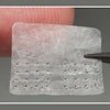
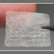
 An intraoral view of an ameloblastoma in the right mandible of an 11-year-old girl. All images courtesy of Bonitz et al. Licensed under CC BY-NC-ND 4.0.
Long road to recovery
During an orthodontic exam in 2011, a clinician noticed a massive, painless cystic lesion on the 11-year-old girl's right horizontal lower jaw and right ascending mandible. The girl experienced no swelling or numbness in her mental or inferior alveolar nerve, the authors wrote.
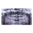
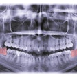
The patient underwent a panoramic x-ray, which revealed extensive bone destruction on her right mandibular angle and the ascendant part through to her collar region. A CT scan revealed that the cyst contained inhomogeneous tissue. Lab tests revealed that the cyst was a unicystic ameloblastoma.
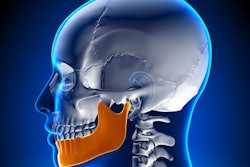
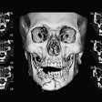
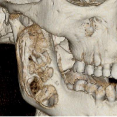
 Reconstruction of CT data shows right mandible.
Ameloblastomas are benign epithelial odontogenic tumors that can derive from enamel residues, parts of the developing enamel, or the basal cells of the oral mucosa, among other tissue. They have the potential to grow very large with resulting bone defects and significant destruction of functional tissue. Unicystic is the least common type of ameloblastoma.
Unicystic ameloblastomas are less aggressive than solid or multicystic ameloblastomas, but they have a strong propensity for recurrence. Therefore, long-term monitoring is necessary for patients who undergo treatment for a unicystic ameloblastoma.
In this case, clinicians resected the ameloblastoma while preserving the lower jaw nerve and the intracapsular condylar. Sections of the inner area of the girl's iliac bones were removed and used to reconstruct her jaw. After the tumor was resected, the iliac bone graft was fixed to the residual condyle, and then the horizontal section of the right jawbone was attached.
Two months after this procedure, the girl underwent a CT scan, revealing that there was no condylar resorption, the authors wrote.
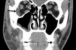
 A CT scan of the girl's jaw two months after surgery.
Four months after the surgery, the internal bone fixation was removed. An x-ray showed that the bone graft had integrated completely, and no iatrogenic osteotomy-line could be seen.
Approximately two years after the ameloblastoma resection and reconstruction, a CT scan showed that the transplanted bone was integrated and remodeled, and the masseter muscle insertion on the mandibular angle was rebuilt, Bonitz and colleagues noted.
 A CT scan of the girl's jaw two years after surgery.
When the patient was 16, clinicians completed a mandibular sagittal split osteotomy to fix her angle class II malocclusion. In February 2017, which was about seven months after the osteotomy, clinicians removed the osteosynthesis from both sides of her mouth and augmented the iliac bone to the molar region of her right mandible and the front maxilla in preparation for placing dental implants. Also, the augmented bone was fixed with screws.
In December 2017, four dental implants were inserted, and in March 2018, crowns were fixed on the implants. In August 2018, the function and esthetic of the girl's mouth was rehabilitated completely, the authors concluded.
Follow-up exams and imaging
Patients who receive ameloblastoma treatment should undergo clinical exams and imaging two times per year for the first five years after the procedure, and once per year after that period. A long-term follow-up period of about 10 years is suggested, they noted.
"A well-thought-out concept of surgical resection and reconstruction can lead to complete rehabilitation," Bonitz and colleagues wrote.


