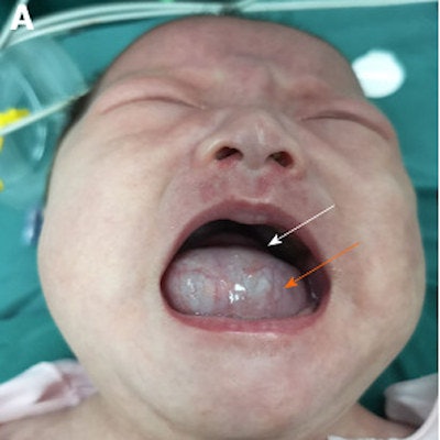
An infant born with multiple rare dermoid cysts in the floor of her mouth, forcing her tongue backward and causing dysphagia, underwent two surgeries before she turned 1 year old due to doctors missing smaller cysts, according to a recent case report.
A magnetic resonance imaging (MRI) scan confirmed a 3-cm cyst, which was first detected during a prenatal exam when her mother was 24 months pregnant, that started at the former midline region in the floor of the girl's mouth. Other lesions were overlooked, likely because doctors were distracted by the more "glaring findings on imaging," according to the case report published in the World Journal of Clinical Cases (July 6, 2020, Vol. 8:13, pp. 2885-2892).
"This report demonstrates the importance of carefully analyzing preoperative imaging to avoid multiple operations for a seemingly isolated oral cyst," wrote the authors, led by Zhi-Ming Wang, DDS, MD, PhD, from the department of stomatology at Shengjing Hospital of China Medical University in Shenyang, China.
Very rare
It is unusual for multiple dermoid cysts to be found in the floor of the mouth. When they do grow in the head or neck, there is often only one mass, and, in most cases, they involve orbital regions.
They are rarely found in infants but may occur at any age. In general, multiple large intraoral dermoid cysts appear in people in their 20s or 30s. Often benign, these cysts are removed via intraoral or extraoral surgery depending on their location.
A 1-month-old baby girl
At week 24 of the mother's pregnancy, the lesion was 1.9 x 1.8 cm. By week 34, it had grown to 2 x 3 cm. At birth, the cyst had grown to nearly fill the patient's oral cavity. Gradually, it affected her breathing and her ability to swallow. At 1 month old, the otherwise healthy girl was taken to the hospital for swelling and dysphasia. The baby could not close her mouth. An MRI revealed a 3.3 x 2.8 x 2.9-cm cystic lesion, pushing the baby's tongue backward, according to the authors.
 The girl prior to her first surgery. A: The white arrow shows her raised tongue in the retracted position. The orange arrow indicates the large-sized cystic mass protruding from her mouth. B: MR imaging reveals a large lesion (orange arrow) and a small suspect lesion (white arrow). C: View in coronal plane of a cystic lesion. D: View in sagittal plane of large (orange arrow) and tiny suspect lesions (white arrow). All images courtesy of Zhi-Ming Wang, DDS, MD, PhD, et al. Licensed under CC BY-NC 4.0.
1st surgery
An intraoral approach was used to remove the cyst due its location near the supramylohyoid muscle. The size of the cyst prevented direct oral intubation, so clear fluid was withdrawn from the mass. She was given general anesthesia, and the cyst was removed. Three days after the surgery, a mild edema developed at the incision site and persisted despite three days of dexamethasone treatment, they wrote.
 The girl during her first surgery. A: Contracted lesion and lowered tongue after clear fluid was aspirated (white arrow at shrunken mass). B: Clear fluid withdrawn. C: Horizontal incision made. D: Dissection of cyst from surrounding tissues. E: Removed cyst. F: Histology revealed keratinizing stratified squamous epithelial lining (white arrows) and sebaceous glands (orange arrows).
 The infant three days after surgery. A: Edema at the incision site. B: Postoperative MRI shows two cystic lesions (orange arrows) under her tongue. C and D: View of each lesion in coronal plane (orange arrow). E: Both lesions in sagittal plane (orange arrows).
A week later, an MRI scan of the tongue revealed two small cystic lesions under the infant's tongue. One was about 0.8 x 1 x 0.5 cm, and the other was 1.2 x 0.5 x 0.5 cm. After reviewing the preoperative MRI, doctors identified a suspicious lesion of minute size that they believe they missed due to the larger cyst compressing it and doctors focusing on the most pronounced findings on the scan. After the sizable cyst was removed, the missed one grew rapidly. Nine days after surgery, the patient was discharged. An exam of the removed cyst showed it was a keratinizing stratified squamous epithelial lining complete with sebaceous adnexa, confirming it was a dermoid cyst, the authors wrote.
2nd surgery
Nine months after surgery, the infant was brought back to the hospital. She had a normal mouth opening but her tongue was slightly raised. The doctor could feel a 2 x 2-cm mass on her tongue muscle near the midline in the floor of her mouth, and an MRI confirmed it. During the surgery to remove this cyst, doctors found another deeply embedded in the mylohyoid muscle at the floor of the mouth, which required careful separation. The content of these lesions was sebum, so they were diagnosed as dermoid cysts.
 The infant during the second surgery. A: Second preoperative MRI of large mass and a tiny suspect lesion (white arrow). B: MRI in sagittal plane showing large high-signal (wide arrow) and tiny suspect (narrow arrow) lesions. C: MRI in coronal plane of one lesion only. D: Completely encapsulated mass. E: Fully removed lesion. F: Cross-section with keratin content.
Five months after the second operation, a 1.5 x 0.9 x 1-cm cyst was found in the same location. Because the lesion was not affecting the girl's eating, swallowing, or breathing, it was not removed. The girl will be closely monitored, the authors wrote.
Origin of the cysts
Because the first cyst was detected during a prenatal exam, the cysts are believed to be congenital. The first and second branchial arches bilaterally join at midline during the early stages of embryonic development. Sometimes, epithelial remnants get trapped or leftovers of odd nodules forming the tongue and floor of the mouth may persist, resulting in dermoid cysts.
Look closely
There are several unusual aspects to this case, including the fact that the dermoid cysts didn't occur as one mass, the condition is rarely found on prenatal exams, and the cysts occurred in the floor of an infant's mouth. Clinicians should take these items into consideration when trying to diagnose patients, they wrote.
"It is of great importance to analyze preoperative imaging carefully and to consider the potential for multiplicity in a seemingly isolated midline cyst of oral cavity," the authors wrote.


