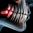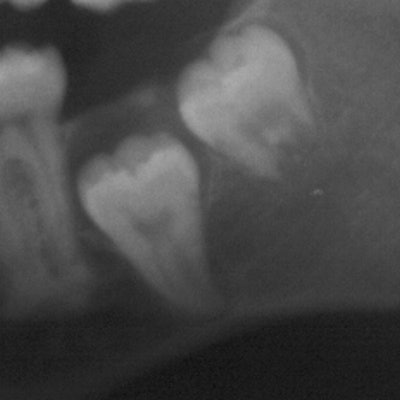
When you are considering extracting an impacted third molar, on what information do you base your decision? The findings of a new study might help you in your next case, as researchers looked at almost 1,200 clinical images to see if a consistent impaction pattern existed and to define the most appropriate age for prophylactic extraction.
They found that patients older than 20 were more likely to have a vertical impaction pattern, while younger patients had a horizontal one. The researchers recommended waiting until a patient is older than 20 to extract these teeth. They published their findings in BMC Oral Health (April 4, 2018).
"Many radiographic readings in patients older than 20 years are in favor of eruption compared with younger patients," the researchers wrote.
The study was led by Soukaina Ryalat, BDS, head of the department of oral and maxillofacial surgery at the University of Jordan Faculty of Dentistry in Amman.
No consensus
The extraction of impacted third molars is one of the most common procedures in oral and maxillofacial surgery. While these teeth are the most likely to be impacted, it is estimated that up to half are removed without clinical or imaging rationale. Why? Because it is difficult to predict if the impacted tooth will cause complications if left in the mouth.
“Many radiographic readings in patients older than 20 years are in favor of eruption compared with younger patients.”
Generally, the question of whether to extract or not is left up to the clinical judgment of the practitioner, the study authors wrote. Some opt for early extraction, while others wait to see if extraction becomes necessary at a later date.
What if there were a way to use imaging technology to determine the most appropriate age for prophylactic extraction of these teeth? The researchers suggested that if the eruption and impaction patterns of third molars could be identified, then they would aid clinical decision-making in deciding whether to extract or not.
The researchers reviewed almost 1,200 panoramic radiographs (Kodak 8000C, Carestream Dental) taken at the University of Jordan Hospital Department of Dentistry between 2010 and 2014. These included 1,810 impacted lower third molars from patients whose ages ranged from 18 to 26. They examined the molars' pattern of eruption in relation to the patients' ages using standard radiographic points and angles.
The researchers defined a third molar's angulation as the angle formed between the dental long axis and occlusal plane:
- Horizontal is defined as less than 20°.
- Mesioangular is defined as between 20° and 80°.
- Vertical was defined as between 80° and 100°.
- Distoangular is defined as 100° or more.
Overall, almost two-thirds of the impactions were mesioangular (66.1%), followed by vertical impactions (18.8%) and horizontal ones (15.1%). The radiographic average width of the impacted molars was 10.1 ± 1.4 mm.
The impaction pattern differed depending on the patient's age, the researchers reported. In those older than age 20, vertical pattern of impaction was the most common (21.4%), but horizontal impaction was more common in younger patients (21.3%).
The third-molar angulation changed significantly between age groups, but the team found no constant pattern from year to year. The angulation of lower third molar teeth was significantly higher in patients older than 20 (46.2° ± 26.8°) compared with younger patients (38.4° ± 25.1°; p < 0.001).
As patients became older, the researchers noted a constant pattern of an increase in the retromolar space (p < 0.001). They measured the retromolar space as the space between the lines of the anterior border of the ramus to the most distal point of the lower second molar. After the age of 25, the space was close to the average width of the lower third molar teeth (see table below). More space could help reduce complications if extraction is undertaken, they noted.
| Measurement of retromolar space by patient age | ||
| Age | No. of impacted third molars | Retromolar space in mm (mean ± standard deviation) |
| 18 | 222 | 40.0 ± 23.4 |
| 19 | 205 | 37.2 ± 24.8 |
| 20 | 208 | 37.8 ± 27.0 |
| 21 | 203 | 49.0 ± 27.7 |
| 22 | 202 | 43.8 ± 28.1 |
| 23 | 217 | 49.8 ± 28.0 |
| 24 | 205 | 45.8 ± 25.5 |
| 25 | 166 | 43.6 ± 24.4 |
| 26 | 182 | 44.2 ± 26.3 |
"This signifies the importance of re-evaluating patient's radiograph considering the possible changes of impacted lower third-molar teeth that could occur with time, especially for younger patients," the authors wrote.
Delaying extraction
As a study limitation, the authors noted that this was a review study. They recommended following patients' clinical and radiological data in a cohort study design or retrospectively in a case-control design for better-quality evidence on this topic.
However, based on their study findings, the researchers concluded that delaying extraction of these teeth may be the better clinical option.
"Late extraction of mandibular third-molar teeth (i.e., after the age of 20) is therefore recommended when prophylactic extraction is considered," the authors wrote.
