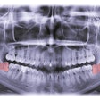
CAD/CAM technology has evolved for use in the dental field since its emergence in the mid-1940s for engineering and design purposes. In dentistry, CAD/CAM technology has been developed for both chairside and laboratory-based applications.
The workflow for creating a computer-generated surgical guide to be used in dental implant surgery is often underutilized because of its traditionally complex workflow. Today, newer technologies are available, such as the Cerec CAD/CAM and Galileos cone-beam CT (CBCT) systems (Sirona), which are designed to integrate with each other and facilitate an improved workflow for computer-guided implantology.
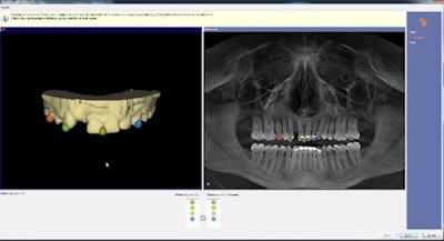 Figure 1: Integration of data between CAD/CAM and CBCT. All images courtesy of Dr. Neal Patel.
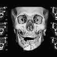
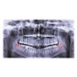
CBCT can be used before a broad range of dental services, including orthodontics, oral surgery, periodontics, endodontics, and prosthodontics, as well as a tool for formulating a comprehensive diagnosis. Sirona's Galileos imaging system takes a 14-second maxillofacial scan and provides 16-cm spherical volume of data.
 Neal Patel, DDS.
In my experience, the CBCT image shows the smallest details in superb clarity while using one of the lowest possible radiation doses among the available units available.
Once the scan has been performed, the images can then be managed within software such as Sidexis 4 (Sirona) and used to aid with treatment planning comprehensive dentistry for a patient.
Patient communication also has become easier. The software streamlines all images in a patient-friendly timeline where they can visualize their dental conditions and compare changes over time. This process has increased the rate of treatment acceptance.
Workflow
The digital implant workflow starts with a diagnostic CBCT scan. Once the scan is obtained, the clinician can evaluate for pathology and evaluate the general anatomy of the area for the future implant. If the patient is a candidate for the implant, a digital CAD/CAM scan is taken. The clinician can design a digital prosthetic wax-up for the patient within the latest software.
With a complete virtual restorative proposal (figure 2), the clinician can send the data to the CBCT software, and the virtual tooth, soft tissue, and bony anatomy can be evaluated for ideal 3D implant planning.
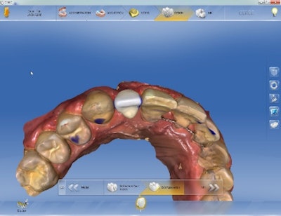 Figure 2: Virtual tooth wax-up.
The major benefits of implant application of CAD/CAM dentistry include improved accuracy and increased fabrication efficiency. Once an ideal implant has been planned (figure 3), the clinician has the choice of several options for guide fabrication. The most popular method includes the digital transfer of data to the SICAT lab in Bonn, Germany, for a lab-grade surgical guide called OptiGuide, complete with master sleeve, drilling protocol, and certificate of accuracy.
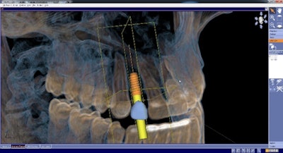 Figure 3: 3D treatment planning of implant and restoration.
The newest option for surgical guide fabrication (figure 4) is called Cerec Guide 2 (Sirona) and allows the clinician to fabricate a surgical guide in house with the MC XL chairside milling unit (Sirona). For this technique, an intraoral scan is taken of the patient's arch with the chairside scanner. A restoration is then virtually created for the missing tooth in a similar manner as a conventional wax-up would be done. Once this virtual tooth wax-up is completed, it is saved as an SSI file, and this file is then imported to the CBCT software.
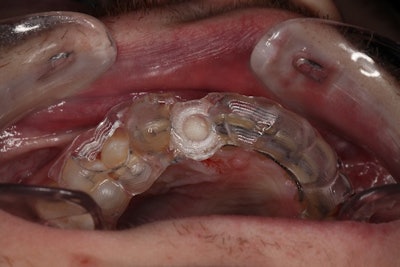 Figure 4: Surgical guide fabricated in office using chairside milling for guided surgery.
A 3D CBCT scan also is taken, which is then integrated with the digital wax-up within the Galileos implant software, allowing the doctor to virtually plan the implant placement in 3D. The digital model can be trimmed during the design of the surgical guide, and parameters can be set for a specific fit and thickness. The automatic proposal of the surgical guide comes with tools available to modify the design before the guide is positioned within a polymethyl methacrylate (PMMA) block and milled using the chairside milling machine within 40 minutes.
Neal Patel, DDS, is an international educator on 3D digital imaging and CBCT treatment planning. He is in private practice in Powell, OH.
Sophia Fratianne serves as editor in chief at Infinite Smiles, Dr. Patel's practice.
The comments and observations expressed herein do not necessarily reflect the opinions of DrBicuspid.com, nor should they be construed as an endorsement or admonishment of any particular idea, vendor, or organization.
