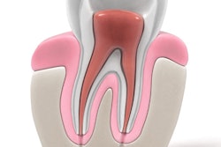
Surprises in root canal therapy, such as additional canals on an incisor, can disrupt the best-laid treatment plans. But can cone-beam CT (CBCT) help?
As it turns out, more than 40% of root canal treatments end up needing retreatment because of missed roots or canals. So researchers from France wanted to determine if the presence of an additional canal on one tooth means an additional canal is likely on another tooth in the same patient.
"The aim of this pilot study was to assess using CBCT acquisitions regarding whether one root canal anatomy of a tooth is associated with a specific anatomy of another tooth," wrote the authors, led by Paul Monsarrat, DDS, PhD, from the department of anatomical sciences and radiology at the Paul Sabatier University Faculty of Dentistry (PLOS One, October 20, 2016).
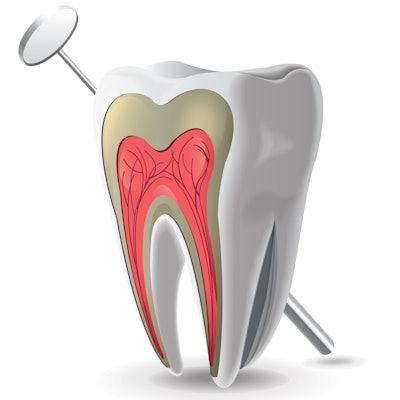
Anatomy of a root canal
Being able to visualize the anatomy of a root canal is critical in endodontic treatments. Successful treatment depends on the practitioner's understanding of a patient's root canal system, according to the study authors. While previous research has studied this anatomy with other imaging techniques, such as electron microscopy and micro-CT, this is the first study that used CBCT to determine if the root canal anatomy of one tooth is associated with a specific anatomy of another tooth in the same patient.
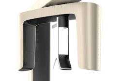
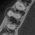
The researchers began with 106 CBCT acquisitions obtained from 2012 to 2013 using a CBCT scanner (CS 9500 3D1, Carestream Dental) with a voxel size of 200 µm. They analyzed more than 2,400 teeth using CS Dental Imaging Software 3DModule v3.2.9.
The age of the patients ranged from 20 to 88 years (mean age of 46.8 years). The study sample included 53 women and 49 men with almost 1,200 maxillary teeth and more than 1,200 mandibular teeth.
In the maxillary incisor-canine group, only slightly more than 1% of teeth had more than one root or canal, the authors reported. However, in mandibular incisor-canine teeth, the number that had two canals in one root jumped to 11% to 13%. They noted that anatomical variations were more frequent in the mandibular central and lateral incisors, where two canals for one root were identified in the following:
“Medium field-of-view acquisitions could be used as an initial database, thus furnishing preliminary evaluations and information.”
- 11% of left central incisors
- 13% of right central incisors
- 12% of left lateral incisors
- 13% of right lateral incisors
In the maxillary first premolars, the researchers found a "large incidence" of teeth with two roots and two canals, observed in 81% to 84% of images. A third root and a third canal were present in fewer than 5% of cases.
The researchers also sought to quantify the chances that CBCT would discover an additional canal on a group of teeth when a variability was found on another group of teeth. When patients had "variability" on a mandibular premolar, they were four times as likely to have variability on mandibular incisors or canines, they noted. They also found that having variability on mandibular incisors or canines increased the risk of having variability on mandibular premolars by four.
However, having variability on mandibular premolars decreased the risk that the practitioner would find variability on mandibular molars with CBCT. Male patients in the study had less than half the risk of having a variability on mandibular molars compared with the female patients, the authors noted.
Resolution issue
The use of CBCT with a resolution of 200 µm was a limitation, as the "coarser resolution" may be responsible for some "uncertainty when describing canal anatomy," the authors reported. They noted that previous studies using different visualization techniques, such as an operating microscope, revealed a higher canal detection rate than CBCT examinations.
"Although CBCT examinations are conducted in the first intention of making a diagnosis or prognostic evaluation, medium field-of-view acquisitions could be used as an initial database, thus furnishing preliminary evaluations and information," the authors concluded.


