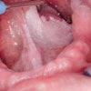
New research by University of Pennsylvania School of Medicine investigators has shown that a protein that helps cells stick together is frequently absent or out of place in squamous cell cancers of the oral cavity and esophagus, but it's unclear if its loss causes the tumors (Cancer Cell, April 12, 2011, Vol. 19:4, pp. 470-483).
The investigators report that mice engineered to lack this protein, called p120 catenin (p120ctn), in the oral-upper digestive tract developed squamous cell cancers. The data could settle a 20-year debate and prove that p120ctn is a tumor-suppressor protein. What's more, the tumors that formed in this mouse model closely resembled human disease and may point the way to novel therapies and early detection strategies.
"As the mice aged, what we saw was a dramatic evolution of precancer to cancer," said senior author Anil Rustgi, MD, a professor and chief of gastroenterology at the school of medicine. "Both the precancerous growth, called dysplasia, and the cancer look exactly like what we see in humans."
In healthy tissues, p120ctn is part of a protein complex that holds epithelial cells in tightly packed sheets. When p120ctn (or another of these cell adhesion proteins) is lost, a variety of cancers, including those in the prostate, breast, pancreas, colon, skin, bladder, and endometrium, can result.
The cells lose their tight cell-cell contacts and can migrate more easily, which likely favors cancer spread and invasion of new cells. However, earlier attempts to test the effects of p120ctn loss on cancer formation were derailed because the animals cannot survive throughout embryonic development or immediately after birth without the protein.
To get around that problem, Dr. Rustgi and colleagues used a system called Cre-Lox that allows them to remove a particular gene in only a subset of tissues. In this case, the team deleted p120ctn from the oral cavity, esophagus, and forestomach. The mutant animals survived through early development and birth, but by 4 to 6 months, most of the mutant animals had developed precancerous lesions and by 9 to 12 months, 70% of the mutant animals had full-blown tumors. By contrast, none of the control littermates developed cancer.
As the lesions developed, the tumor cells secreted inflammatory signals that acted like homing signals for immune cells called immature myeloid cells, the investigators noted. These cells, in turn, helped to create a microenvironment that supported tumor growth. In fact, when the researchers blocked recruitment of the immune cells, they saw a dramatic reduction in tumor invasion.
Based on these observations, Dr. Rustgi said he thinks that targeting the immature myeloid cells may reverse or slow tumor grown in humans, although more work needs to be done in animal models before the approach is tested in the clinic.